

SARS-CoV-2 polyprotein substrate regulates the stepwise Mpro cleavage reaction
- PMID: 37044215
- PMCID: PMC10084705
- DOI: 10.1016/j.jbc.2023.104697
SARS-CoV-2 polyprotein substrate regulates the stepwise Mpro cleavage reaction
Abstract
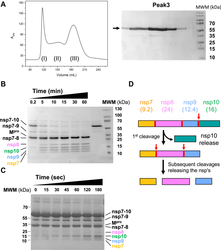
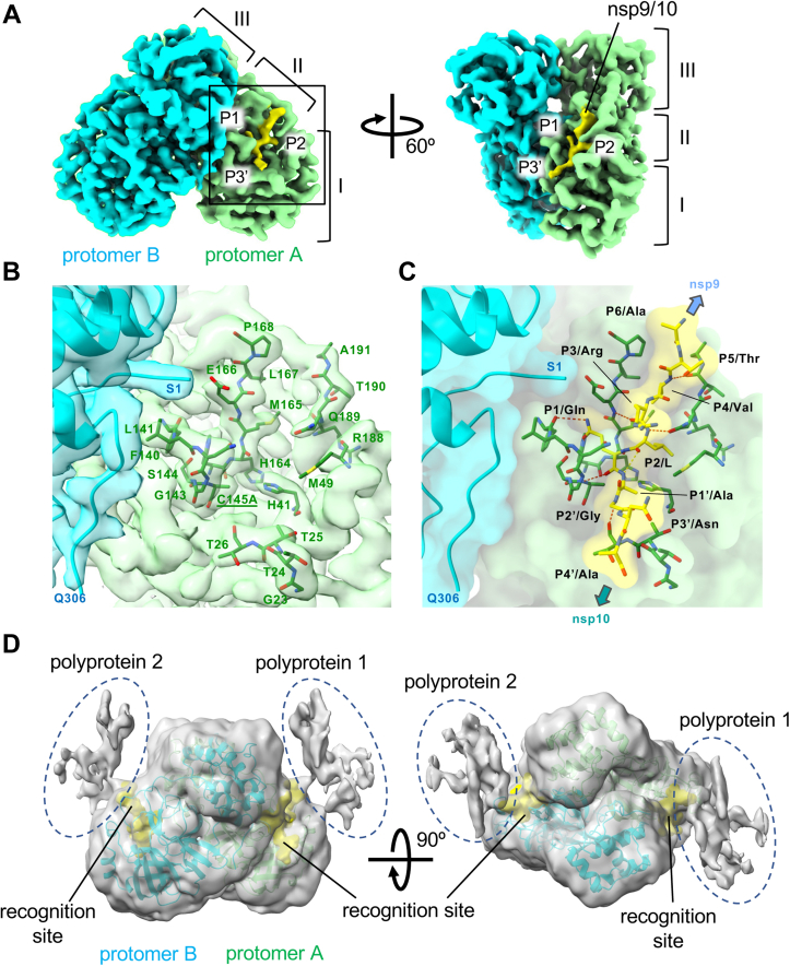
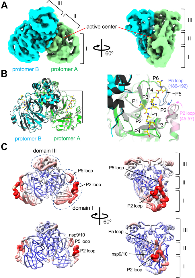
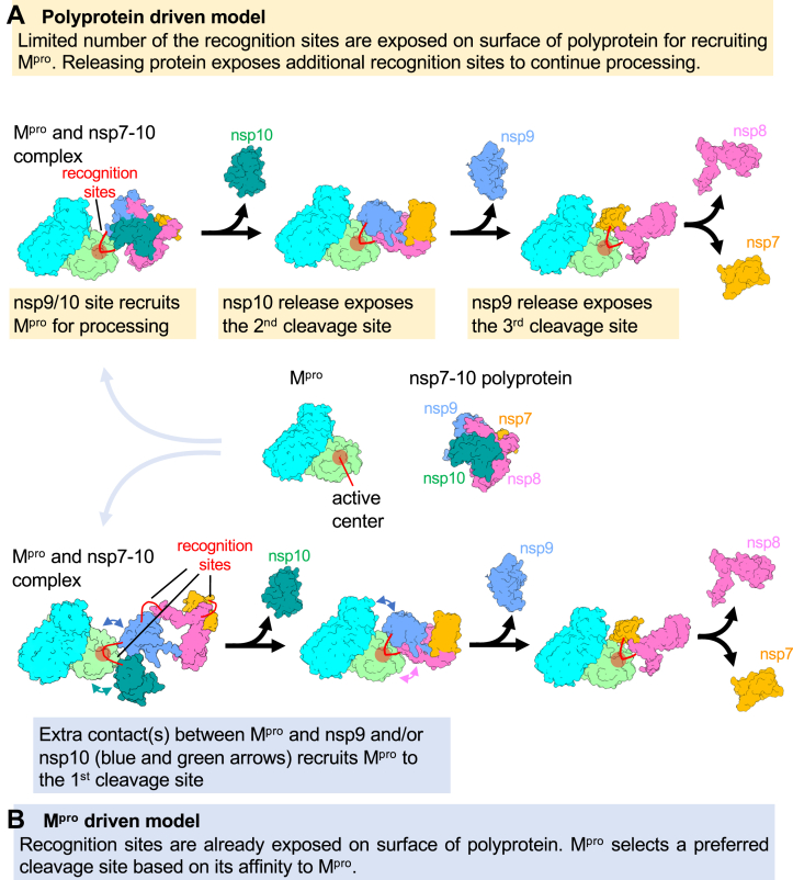
The processing of the Coronavirus polyproteins pp1a and pp1ab by the main protease Mpro to produce mature proteins is a crucial event in virus replication and a promising target for antiviral drug development. Mpro cleaves polyproteins in a defined order, but how Mpro and/or the polyproteins determine the order of cleavage remains enigmatic due to a lack of structural information about polyprotein-bound Mpro. Here, we present the cryo-EM structures of SARS-CoV-2 Mpro in an apo form and in complex with the nsp7-10 region of the pp1a polyprotein. The complex structure shows that Mpro interacts with only the recognition site residues between nsp9 and nsp10, without any association with the rest of the polyprotein. Comparison between the apo form and polyprotein-bound structures of Mpro highlights the flexible nature of the active site region of Mpro, which allows it to accommodate ten recognition sites found in the polyprotein. These observations suggest that the role of Mpro in selecting a preferred cleavage site is limited and underscores the roles of the structure, conformation, and/or dynamics of the polyproteins in determining the sequence of polyprotein cleavage by Mpro.
Keywords: 3CL main protease (M(pro)); SARS CoV-2; cryogenic electron microscopy (cryo-EM); polyprotein; proteolytic processing.
Copyright © 2023 The Authors. Published by Elsevier Inc. All rights reserved.
Conflict of interest statement
Conflict of interest The authors declare that they have no known competing financial interests or personal relationships that could have appeared to influence the work reported in this paper.
Figures
Similar articles
-
Kinetic comparison of all eleven viral polyprotein cleavage site processing events by SARS-CoV-2 main protease using a linked protein FRET platform.J Biol Chem. 2024 Jun;300(6):107367. doi: 10.1016/j.jbc.2024.107367. Epub 2024 May 15. J Biol Chem. 2024. PMID: 38750796 Free PMC article.
-
Potential 3-chymotrypsin-like cysteine protease cleavage sites in the coronavirus polyproteins pp1a and pp1ab and their possible relevance to COVID-19 vaccine and drug development.FASEB J. 2021 May;35(5):e21573. doi: 10.1096/fj.202100280RR. FASEB J. 2021. PMID: 33913206 Free PMC article. Review.
-
Three-dimensional domain swapping as a mechanism to lock the active conformation in a super-active octamer of SARS-CoV main protease.Protein Cell. 2010 Apr;1(4):371-383. doi: 10.1007/s13238-010-0044-8. Epub 2010 May 8. Protein Cell. 2010. PMID: 21203949 Free PMC article.
-
Characterization of Self-Processing Activities and Substrate Specificities of Porcine Torovirus 3C-Like Protease.J Virol. 2020 Sep 29;94(20):e01282-20. doi: 10.1128/JVI.01282-20. Print 2020 Sep 29. J Virol. 2020. PMID: 32727876 Free PMC article.
-
Spatial and temporal roles of SARS-CoV PLpro -A snapshot.FASEB J. 2021 Jan;35(1):e21197. doi: 10.1096/fj.202002271. FASEB J. 2021. PMID: 33368679 Free PMC article. Review.
Cited by
-
Molecular Recognition of SARS-CoV-2 Mpro Inhibitors: Insights from Cheminformatics and Quantum Chemistry.Molecules. 2025 May 15;30(10):2174. doi: 10.3390/molecules30102174. Molecules. 2025. PMID: 40430346 Free PMC article.
-
Recognition and cleavage of human tRNA methyltransferase TRMT1 by the SARS-CoV-2 main protease.Elife. 2025 Jan 7;12:RP91168. doi: 10.7554/eLife.91168. Elife. 2025. PMID: 39773525 Free PMC article.
-
High-Resolution Substrate Specificity Profiling of SARS-CoV-2 Mpro; Comparison to SARS-CoV Mpro.ACS Chem Biol. 2024 Jul 19;19(7):1474-1483. doi: 10.1021/acschembio.4c00096. Epub 2024 Jun 12. ACS Chem Biol. 2024. PMID: 38865301 Free PMC article.
-
Distal protein-protein interactions contribute to nirmatrelvir resistance.Nat Commun. 2025 Feb 1;16(1):1266. doi: 10.1038/s41467-025-56651-x. Nat Commun. 2025. PMID: 39893201 Free PMC article.
-
The zymogenic form of SARS-CoV-2 main protease: A discrete target for drug discovery.J Biol Chem. 2025 Jan;301(1):108079. doi: 10.1016/j.jbc.2024.108079. Epub 2024 Dec 14. J Biol Chem. 2025. PMID: 39675720 Free PMC article.
References
-
- Gupta S.P. Elsevier/Academic Press; London: 2017. Viral Proteases and Their Inhibitors.
Publication types
MeSH terms
Substances
Grants and funding
LinkOut - more resources
Full Text Sources
Miscellaneous
