. 2023 Jul 15;63(7):265-272.
doi: 10.2176/jns-nmc.2022-0283. Epub 2023 Apr 13.

A Consideration of Optimal Head Position in Transsylvian Selective Amygdalohippocampectomy

Affiliations

A Consideration of Optimal Head Position in Transsylvian Selective Amygdalohippocampectomy

Tadashi Hamasaki et al. Neurol Med Chir (Tokyo). .

Abstract

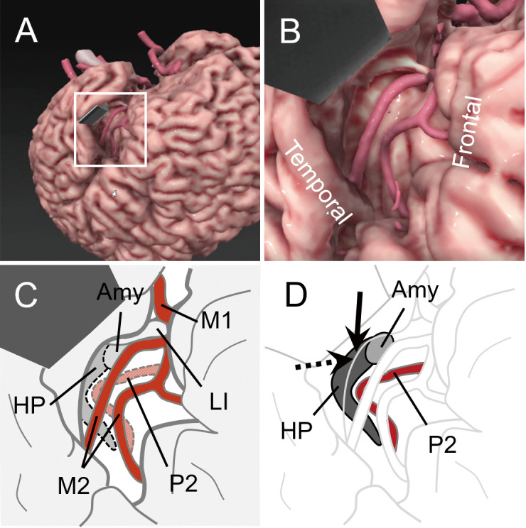
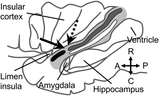
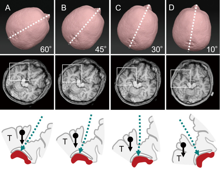
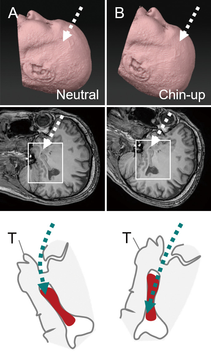
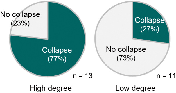
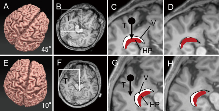
Transsylvian selective amygdalohippocampectomy (TSA) is one of the predominant surgical options for drug-resistant mesial temporal lobe epilepsy. The purpose of this article is to highlight the unique features of TSA and determine the setting to perform safe and secure TSA with special reference to the optimal head position. TSA should be performed via a small surgical corridor in the temporal stem that contains functionally important fiber tracts, including the uncinate fasciculus, the inferior fronto-occipital fasciculus, and the optic radiation. Graphical simulations proposed that low-degree (<30°) head rotation had the advantage of sufficiently opening the surgical field in TSA and may help surgical procedures within the limited exposure of the medial temporal structures. Inspection of the surgical videos implied that the collapse of the inferior horn was prevented in low-degree rotation, probably because the deformation due to the brain shift was minimized in the medial temporal structures. A simulation also implied that chin-up position had the advantage of resecting the tail of the hippocampus in a straightforward manner. We suggest that the setting is optimized in TSA with low-degree rotation and chin-up head position.

Keywords: amygdalohippocampectomy; anterior temporal lobectomy; head position; lateral ventricle; mesial temporal lobe epilepsy.


Conflict of interest statement

The authors declare no conflict of interest related to this work.

Figures

Fig. 1
Fig. 1
Graphical computer simulation and schematic diagrams of the surgical field in transsylvian selective amygdalohippocampectomy. A. A macroscopic view after opening the left Sylvian fissure. The temporal lobe is retracted using a spatula. The boxed area is enlarged in B. The image was generated using software specialized for surgical simulation (GRID, Kompath Inc., Tokyo). B. An enlarged view of A. In the center, two branches of the middle cerebral artery (MCA) are exposed on the insular cortex. C. A schematic diagram of C. The M1 and M2 portions of the MCA are indicated. Dotted lines delineate the locations assumed for the hippocampus (HP), amygdala (Amy), and P2 portion of the posterior cerebral artery in the deeper layer. LI, the limen insulae. D. The same view as C, in which HP, Amy, and P2 are illuminated. An arrow indicates the cortical incision on the inferior periinsular sulcus to reach the inferior horn of the lateral ventricle and the HP. A dotted arrow indicates the trajectory to resect the amygdala.
Fig. 2
Fig. 2
Fiber tracts in the temporal stem. A lateral view of the left temporal stem (tiny dotted area) after removing the frontal and temporal operculi. The black, gray, and white bands indicate the uncinate fasciculus (UF), inferior fronto-occipital fasciculus (IFOF), and optic radiation (OR), respectively. The hippocampus is seen through a window opened on the inferior horn of the lateral ventricle (ventricle). The arrow indicates the surgical corridor in the temporal stem made to reach the inferior horn and hippocampus via a putative junction between the UF and IFOF/OR. A dotted arrow indicates the trajectory to resect the amygdala. The diagram was drawn according to references from Kier et al.) and Ribas et al.)
Fig. 3
Fig. 3
Degree of head rotation and surgical fields on the axial plain. Schematic diagrams showing cases with 60° (A), 45° (B), 30° (C), and 10° (D) head rotations to the unoperated side (upper row). The boxed area in the axial magnetic resonance image, including the Sylvian fissure and hippocampus (middle row), is enlarged and schematized (bottom row). An arrow with a dot indicates the gravity to the temporal lobe (T). The hippocampus is drawn as a (red) solid object at the bottom. The direction of the surgical trajectory is indicated by the (green) dashed lines. Note that the direction of gravity to the temporal lobe is more overlapped with the surgical trajectory at higher degrees of head rotation.
Fig. 4
Fig. 4
Chin-up position and surgical fields on the sagittal plain. Schematic diagrams showing cases in the neutral (A) and chin-up (B) positions (upper row). The boxed area in the sagittal MR image, including the Sylvian fissure and hippocampus (middle row), is enlarged and schematized (bottom row). The hippocampus is drawn as a (red) solid object. The direction of the surgical trajectory is indicated by the (green) dashed lines. Note that the long axis of the hippocampus is on the straight surgical trajectory.
Fig. 5
Fig. 5
Collapse of the inferior horn of the lateral ventricle. Collapse of the inferior horn is evaluated in video recordings of 24 transsylvian selective amygdalohippocampectomy surgeries. The rates of collapse are 77% (10/13) and 27% (3/11) in high (30° or more) and low (less than 30°) degrees of head rotation, respectively. The difference is statistically significant at p < 0.05 (chi-square test).
Fig. 6
Fig. 6
Degree of rotation, brain shift, and collapse of the ventricle. A-D. A simulation of 45° head rotation. A boxed area in an axial slice of MRI (B) is enlarged in C. An arrow with a dot in C indicates the gravity to the temporal lobe. A curbed white area and (red) solid object indicate the inferior horn of the lateral ventricle (V) and the hippocampus (HP), respectively. Note that the V and HP reside directly beneath the temporal lobe (T). In D, the space of the ventricle collapses because the temporal lobe has sunken toward the ventricle because of the brain shift during Sylvian fissure opening. E-H. A simulation of 10° head rotation. The collapse of the ventricle is minimum in H because the direction of the brain shift (an arrow with a dot) of the temporal lobe (T) is outside the area containing the ventricle (V) and the hippocampus (HP).

References

    1. Yaşargil MG, Teddy PJ, Roth P: Selective Amygdalo-hippocampectomy: operative anatomy and surgical technique. In: Advances and Technical Standards in Neurosurgery. Vol 12. Symon L, Brihaye J, Guidetti B, et al. , Eds. Springer-Verlag, Wien, New York, 1985: 93-123 - PubMed
    1. Morino M: Selective amygdalohippocampectomy. In: Microsurgery of Intractable Epilepsy. Morino M, Ed. Medical View Co., Ltd., Tokyo, 2013: 16-26
    1. Yonekawa Y: [Operative neurosurgery: personal view and historical backgrounds (4). Selective amygdalohippocampectomy SAHE]. No Shinkei Geka 35: 1183-1196, 2007(Japanese) - PubMed
    1. Von Der Heide RJ, Skipper LM, Klobusicky E, Olson IR: Dissecting the uncinate fasciculus: disorders, controversies and a hypothesis. Brain 136: 1692-1707, 2013 - PMC - PubMed
    1. Helmstaedter C, Richter S, Roske S, Oltmanns F, Schramm J, Lehmann TN: Differential effects of temporal pole resection with amygdalohippocampectomy versus selective amygdalohippocampectomy on material-specific memory in patients with mesial temporal lobe epilepsy. Epilepsia 49: 88-97, 2008 - PubMed