. 2023 Apr 12;14(1):2075.
doi: 10.1038/s41467-023-37726-z.

Collagen breaks at weak sacrificial bonds taming its mechanoradicals

Affiliations

Collagen breaks at weak sacrificial bonds taming its mechanoradicals

Benedikt Rennekamp et al. Nat Commun. .

Abstract

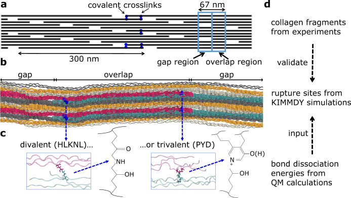
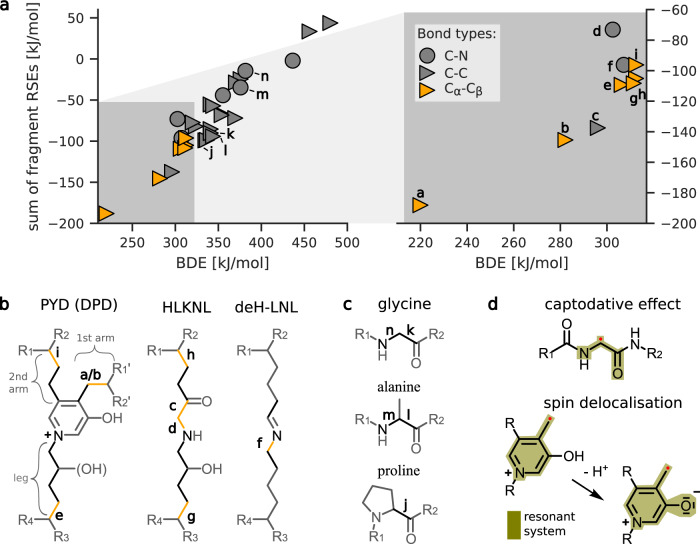
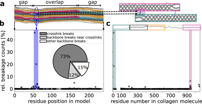
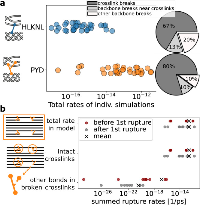
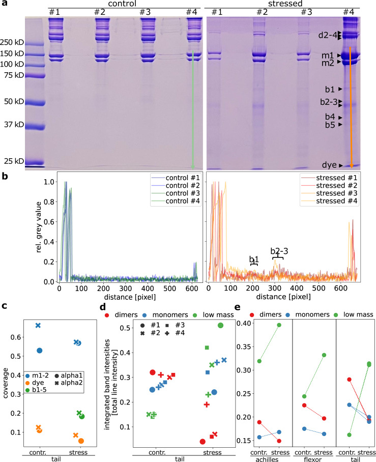
Collagen is a force-bearing, hierarchical structural protein important to all connective tissue. In tendon collagen, high load even below macroscopic failure level creates mechanoradicals by homolytic bond scission, similar to polymers. The location and type of initial rupture sites critically decide on both the mechanical and chemical impact of these micro-ruptures on the tissue, but are yet to be explored. We here use scale-bridging simulations supported by gel electrophoresis and mass spectrometry to determine breakage points in collagen. We find collagen crosslinks, as opposed to the backbone, to harbor the weakest bonds, with one particular bond in trivalent crosslinks as the most dominant rupture site. We identify this bond as sacrificial, rupturing prior to other bonds while maintaining the material's integrity. Also, collagen's weak bonds funnel ruptures such that the potentially harmful mechanoradicals are readily stabilized. Our results suggest this unique failure mode of collagen to be tailored towards combatting an early onset of macroscopic failure and material ageing.


Conflict of interest statement

The authors declare no competing interests.

Figures

Fig. 1
Fig. 1. Collagen structure, spanning multiple length scales, and our corresponding methods.
a 2D projection of the staggered arrangement of collagen triple helices, each 300 nm. This results in the typical overlap and gap regions of collagen, including the 3D braiding of triple helices. b Our atomistic model spanning one overlap (middle) and about one gap region (split into two parts) of collagen. c Zoom in on enzymatic crosslinks connecting the triple helices. Different chemistries (divalent or trivalent) are possible at these positions, for example, Hydroxylysino-keto-norleucine (HLKNL) or Pyridinoline (PYD). d Our workflow combines different methods as collagen spans multiple length scales.
Fig. 2
Fig. 2. Crosslinks harbor the weakest bonds in collagen type I.
a Correlation between the bond dissociation energies (BDEs) and summed radical stabilization energies (RSEs) of homolytically cleaved bonds and formed radicals. All molecular structures used for calculations can be found in the SI. Bonds with BDEs <315 kJ mol−1 are shown in more detail on the right. The small letters close to selected data points indicate respective bonds in the lower panels b and c. b, c Molecular structures of (b) investigated collagen crosslinks and (c) prevalent aminoacids in collagen. BDEs of bonds colored in black and orange (BDE <315 kJ mol−1) were explicitly calculated. Residues R1, R1', R2 and R2' connect to the same collagen triple helix. The BDEs of the PYD bond denoted with the letter “a” was obtained after deprotonation of the pyridine-bound hydroxyl group. d Main electronic effects leading to the lowest found BDEs. The captodative effect describes the non-additive stabilization of the amino-carbonyl and carbonyl-amino group present at any Cα peptide radical. The lower part illustrates the increased radical delocalization upon deprotonation of PYD and DPD, lowering the BDE and RSE of bond “b” to that of bond “a”.
Fig. 3
Fig. 3. Collagen ruptures primarily in crosslinks, but also unspecific ruptures occur everywhere.
Data were obtained from 63 independent MD simulations, including divalent and trivalent crosslink setups, with different collagen sequences and pulling conditions, as described in the methods section. For simulations with trivalent crosslinks, we used the deprotonated BDEs of PYD. In both panels b and c the same data is presented in two different reference systems. a Left side: Location of residues and crosslinks in our model along the fiber axis. Triple helices with the same phase are in the same color, respectively. Upper and lower residues participating in the same crosslinks (blue) appear in the same residue position in the model; see, for example, the blue encircled area. This view is used in panel b. Right side: Crosslinks along a whole 300 nm collagen molecule occur at the beginning (cyan area) and at the end of a triple helix (magenta area), shown as a cartoon with coloring as in the structure on the left. This reference system, counting along the collagen molecule, is used in panel c. b Propensity of bond breakages in our collagen model. Most ruptures concentrate in the crosslinked area, while there are some scattered backbone ruptures. Inset: Pie chart of summed-up ruptures in the crosslinks vs. the backbones in the crosslinked area (up to five residues before/behind) vs. elsewhere in the backbone. c Propensity of bond breakages within collagen fibrils. The crosslinked areas at the beginning and end of the molecules, as marked exemplary for the N-terminal crosslink in the shaded areas, are most prone to rupture. Note that both in b and c we only show data points where at least one rupture happened.
Fig. 4
Fig. 4. Trivalent crosslinks break faster, but (at first) without loosing connection.
a Comparison of total primary bond rupture rates in simulations with models having divalent (HLKNL) vs. models having trivalent (PYD) crosslinks, which show overall higher rates. The ratio of crosslink ruptures to other breakages confirms that the increased rupture rates are indeed due to higher propensities in the crosslinks. b Comparison of secondary bond rupture rates before and after the first arm in one PYD crosslink broke. The rate in the second arm even decreases, being several orders of magnitude lower compared to the sum in the remaining model. Hence, other bonds will break first and the fibril stays overall intact. Each data point is from one of nine simulations with trivalent crosslinks, which we continued after the first rupture in the short PYD arm.
Fig. 5
Fig. 5. Experiments support the simulations of covalent backbone and crosslink rupture in stressed tendon collagen.
a SDS-PAGE analysis reveals altered patterns of bands and their intensities between stressed and control tendon collagen. The four lanes are technical repeats, the experiment was repeated independently for a second biological sample set, see Supplementary Fig. 10. We indicate lines used for analysis in panel b in lane #4. The bands b1–5, cut out for the mass spectrometry, are marked with arrows. b Densitometry to quantify stained collagen α-chains and degradation products on SDS-PAGE. We show the normalized inverted gray value along the lanes. c Percent sequence coverage of collagen α1(I)- and α2(I)-chains from the bands analyzed by mass spectrometry. Collagen fragments are present in the mass regime of monomers m1 and m2, low mass bands b1–b5 (averaged, individual data in Supplementary Fig. 9) and even the dye front. d Integrated intensity in the two monomeric bands (blue) and three dimer (red) bands, as well as the total area below the monomers (incl. dye front, green), normalized for each lane with the total intensity. e Same analysis as in panel d for additional gels provided in Supplementary Fig. 10 from Rat tail, flexor, and achilles tendons.

Similar articles

Cited by

References

    1. Komi PV. Relevance of in vivo force measurements to human biomechanics. J. Biomech. 1990;23:27–34. doi: 10.1016/0021-9290(90)90038-5. - DOI - PubMed
    1. Zapp, C. et al. Mechanoradicals in tensed tendon collagen as a source of oxidative stress. Nat. Commun. 11, 2315 (2020). - PMC - PubMed
    1. Staudinger E, Leupold H. Über Isopren und Kautschuk, 18. Mitteil.: Viscosittäts-Untersuchungen an Balata. Ber Dtsch Chem Ges. 1930;63:730–733. doi: 10.1002/cber.19300630329. - DOI
    1. Caruso MM, et al. Mechanically-induced chemical changes in polymeric materials. Chem. Rev. 2009;109:5755–98. doi: 10.1021/cr9001353. - DOI - PubMed
    1. Luo, Y.-R. Comprehensive Handbook of Chemical Bond Energies (CRC Press, 2007).

Publication types