Clinical Trial
. 2023 Apr 12;14(1):2064.
doi: 10.1038/s41467-023-37845-7.

A case study of percutaneous epidural stimulation to enable motor control in two men after spinal cord injury

Affiliations
Clinical Trial

A case study of percutaneous epidural stimulation to enable motor control in two men after spinal cord injury

Ashraf S Gorgey et al. Nat Commun. .

Abstract

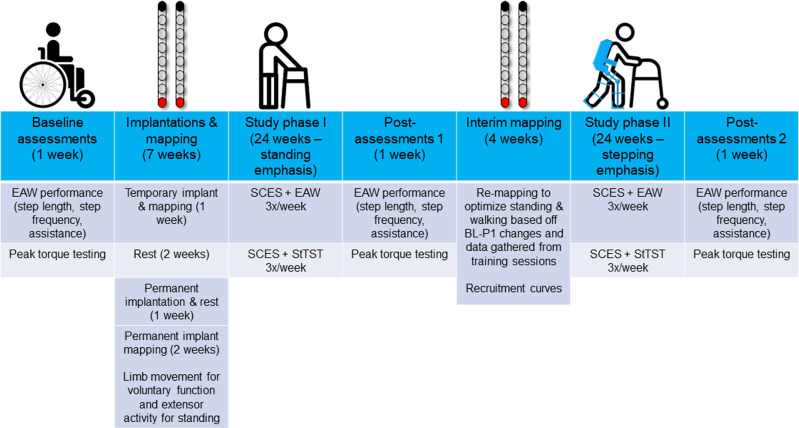
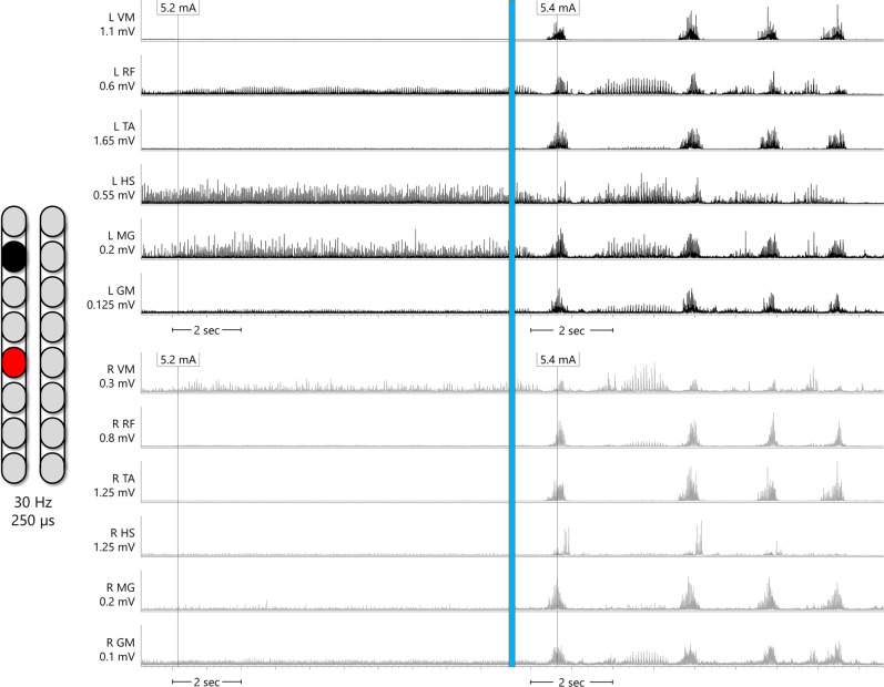
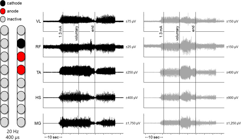
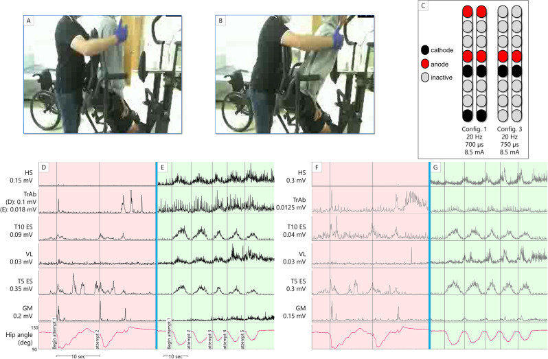
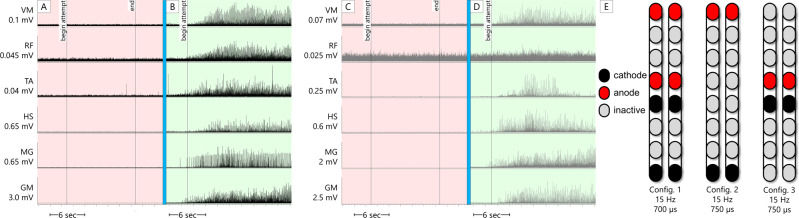
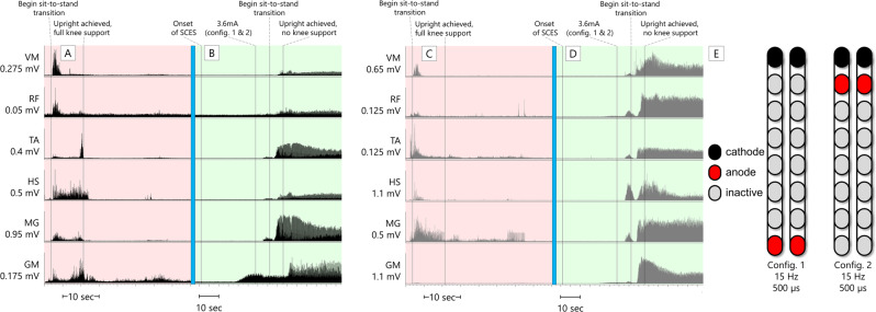
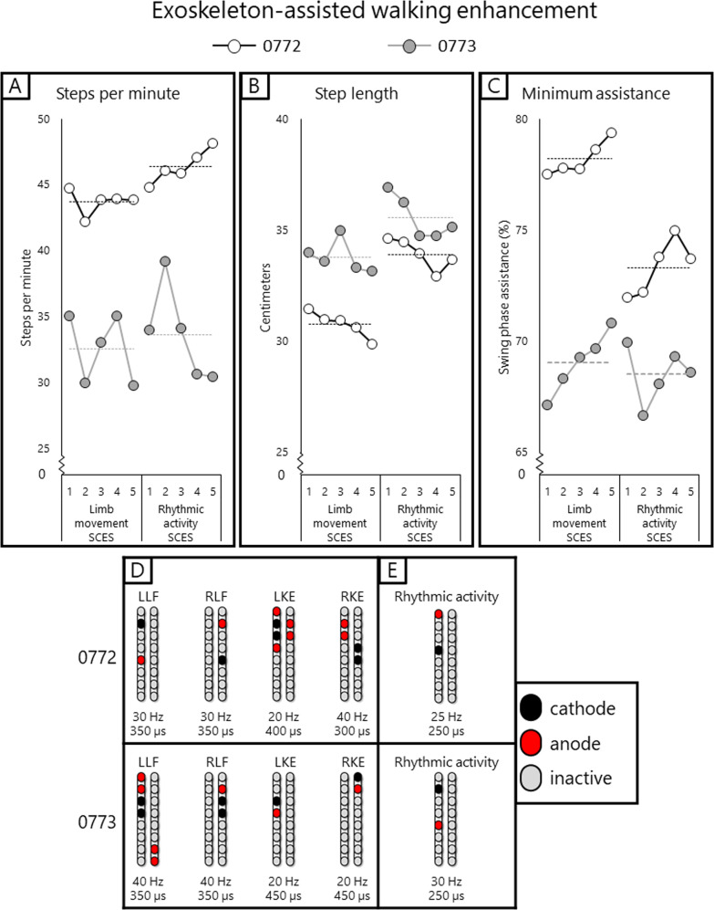
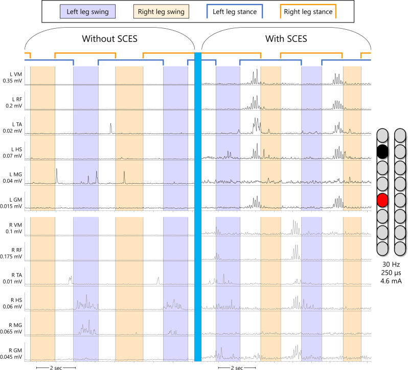
Two persons with chronic motor complete spinal cord injury (SCI) were implanted with percutaneous spinal cord epidural stimulation (SCES) leads to enable motor control below the injury level (NCT04782947). Through a period of temporary followed by permanent SCES implantation, spinal mapping was conducted primarily to optimize configurations enabling volitional control of movement and training of standing and stepping as a secondary outcome. In both participants, SCES enabled voluntary increased muscle activation and movement below the injury and decreased assistance during exoskeleton-assisted walking. After permanent implantation, both participants voluntarily modulated induced torques but not always in the intended directions. In one participant, percutaneous SCES enabled motor control below the injury one-day following temporary implantation as confirmed by electromyography. The same participant achieved independent standing with minimal upper extremity self-balance assistance, independent stepping in parallel bars and overground ambulation with a walker. SCES via percutaneous leads holds promise for enhancing rehabilitation and enabling motor functions for people with SCI.


Conflict of interest statement

The authors declare no competing interests.

Figures

Fig. 1
Fig. 1. Study timeline.
Timeline of study phases for NCT04782947. After baseline outcome measures are assessed, temporary implantation occurs followed by 5 days of mapping. After removal of the temporary implant, participants are given 2 weeks rest and then receive the permanent implant. Permanent implant mapping occurs for 2 weeks, focusing mainly on voluntary limb movement and extensor activity to facilitate standing. Following this, phase I of the study begins, consisting of 24 weeks training 3 days per week. Each training day consists of one hour of spinal cord epidural stimulation (SCES) combined with exoskeleton-assisted walking (EAW), followed by one hour of SCES combined with standing task-specific training (StTST). After 1 week of re-assessing outcome measures (Post-assessments 1), 4 weeks of re-mapping is conducted to optimize standing, and walking function for the next phases of the study. Following this, phase II of the study begins, consisting of 24 weeks of the same training as phase I. In the final week of the study, outcome measures are re-assessed a final time (post-assessments 2).
Fig. 2
Fig. 2. Supine rhythmic EMGs.
Example electromyograms (EMGs) of left leg muscles (black traces) and right leg muscles (gray traces) while SCES is delivered in supine at various amplitudes to participant 0773, using the configuration shown on the left of the figure (black is the cathode and the red is the anode). In the left column of EMGs, SCES delivered at 5.2 mA induces little tonic activity or no activity at all. In the right column of EMGs, the same SCES configuration is delivered at 5.4 mA. Upon ramping of stimulation to 5.4 mA, a single burst of activity across all muscles is induced, followed by periods of relaxation and then subsequent bursts of similar shape. Each burst of muscle activity across all muscles resulted in sudden, bilateral knee, hip and ankle flexion, followed by the legs returning to resting on the table during periods of relaxation. EMGs presented are rectified and bandpass filtered at 10–990 Hz. L left, R right, VM vastus medialis, RF rectus femoris, TA tibialis anterior, HS hamstring, MG medial gastrocnemius, GM gluteus medius, mV millivolts, mA milliamps, Hz hertz, µs microseconds, sec seconds.
Fig. 3
Fig. 3. Configuration for 0773 resulting in right leg flexion.
Left leg muscles are depicted in black and right leg muscles in gray. Both plots occurred simultaneously but have been separated to better visualize different effects of voluntary effort between legs. At 1.3 mA, bilateral muscle activity was induced. However, when the participant volitionally attempted to flex his right leg, muscle activity decreased in some left leg muscles (VL, RF, and HS) while increasing or bursting in right leg muscles (RF, TA, HS, and MG). Hz hertz (stimulation frequency), µs microseconds, L left, R right, VL vastus lateralis, RF rectus femoris, TA tibialis anterior, HS hamstrings, MG medial gastrocnemius, sec seconds, µv microvolts.
Fig. 4
Fig. 4. Voluntary hip flexion and extension with trunk control (participant 0772) from a semi-standing position in a standing frame.
Panels A and B show the start and end of the range of motion, respectively, the participant attempts without self-assistance from the arms or external assistance. A study team member is seen guarding the participant for safety. C SCES configurations and stimulation parameters. DG root mean square EMG activation of trunk (T5 ES and T10 ES: erector spinae muscles at T5 or T10 vertebral levels respectively; TrAb: transverse abdominis muscles) and leg muscles. Trunk EMGs had an additional comb filter applied to remove SCES artifact, while ECG artifact is still visible. Hip angle is derived from an electrogoniometer on the participant’s hip. Panels D and F show left and right-side muscles, respectively, with the hip angle trace replicated in both panels, while the participant attempts the maneuver with SCES off. Bursts seen in ES and gluteal muscles (GM) are likely reflexive, as they occur synchronously with the rapid decline in hip angle as the participant was unable to control his descent. Following this, activity in T5 ES coincides with the participant using arm self-assistance to return to the starting position. Panels E and G show left and right muscles, respectively, with the hip angle trace replicated in both panels, while the participant attempts the maneuver with SCES on. The hip angle shows more controlled descent into flexion and ascent into extension, accomplished without self-assistance or external assistance. EMG activity shown corresponds to hip angle changes – at the lowest hip angle, the participant had the greatest need for trunk stability, and more activity is seen from trunk muscles. Quadriceps and hamstrings increase activity as the participant leans forward, aiding in control of the descent and subsequent ascent. Note that for the left TrAb, y-axis scales are different between panels D and E, in order to better illustrate change in EMG activity during the maneuver with SCES on. HS hamstrings, VL vastus lateralis, GM gluteus medius, sec seconds, config configuration, Hz Hertz, µs microseconds, mA milliamps.
Fig. 5
Fig. 5. 0772 Exoskeleton sit-to-stands.
Electromyograms (EMGs) of lower extremity muscles of 0772 during active attempts to complete a sit-to-stand maneuver with the exoskeleton in “Squat” mode. In this mode, the exoskeleton will not passively complete a sit-to-stand for the user; rather, assistive torque is provided at the knees and hips which can enable the user to complete a sit-to-stand only if they are able to volitionally contribute to the movement. The assistance level was set to “very high”, which is the highest of four levels of manufacturer-determined assistance. A, C left (black traces) and right (gray traces) leg muscles, respectively, during an active attempt to complete a sit-to-stand with SCES off. 0772 was unable to generate any muscle activity, and consequently, could not complete the sit-to-stand. B, D left (black traces) and right (gray traces) leg muscles, respectively, during an active attempt to complete a sit-to-stand with SCES on at 4.2 mA using configurations shown in E. As the muscle activity increased, 0772 was successfully able to complete the sit-to-stand. EMGs presented are rectified and band-pass filtered at 10–990 Hz. VM vastus medialis, RF rectus femoris, TA tibialis anterior, HS hamstrings, MG medial gastrocnemius, GM gluteus medius, sec seconds, Hz Hertz, µs microseconds, config configuration.
Fig. 6
Fig. 6. 0773 overground standing.
Electromyograms (EMGs) of lower extremity muscles of 0773 during overground standing in parallel bars. A, C left (black traces) and right (gray traces) leg muscles, respectively, during upright, overground standing with SCES off. 0773 was able to use his upper extremities to self-assist himself into an upright position with the knees blocked by a study team member, and once upright, required full support at the knees to maintain the upright position. EMG activity during the sit-to-stand transition was a brief period of spasticity – once upright, muscle activity largely ceased. B, D left (black traces) and right (gray traces) leg muscles, respectively, during upright, overground standing with SCES on using configurations shown in E. From the onset of SCES, the amplitude was gradually ramped up on one configuration at a time before both configurations were set at an amplitude of 3.6 mA. After using upper extremities to self-assist into an upright position with the knees blocked by a study team member, the study team member was able to fully let go of the knees. The participant needed no support to maintain standing aside from balance self-assist with the upper extremities. EMGs presented are rectified and band-pass filtered at 10–990 Hz. VM vastus medialis, RF rectus femoris, TA tibialis anterior, HS hamstrings, MG medial gastrocnemius, GM gluteus medius, sec seconds, Hz hertz, µs microseconds, config configuration.
Fig. 7
Fig. 7. EAW enhancement figure.
Data points showing changes in exoskeleton-assisted walking (EAW) performance for two participants walking in “Adaptive” mode, a manufacturer setting which adjusts the amount of assistance provided to the user, and thus can result in variability in certain parameters of gait. AC individual dots represent the average of 300 consecutive steps across 5 separate EAW training sessions for steps per minute, step length, or minimum exoskeleton-provided swing phase assistance (measured as a percentage of the maximum possible assistance the exoskeleton can provide), respectively. Dotted lines represent the average of each 5-session segment. The “Limb movement SCES” segment of each graph show the individual session data (dots and solid lines) and 5-session average (dotted lines) of the last five EAW sessions using four concurrent SCES programs which facilitated flexion or extension movements for each individual leg (configurations shown in panel D). These data are from the last five sessions of a 24-week period of EAW using these configurations. After mapping for supine rhythmic locomotor activity (configurations shown in panel E), improvements are seen in all outcomes for both participants across the first five EAW sessions using this new, single configuration. Increases in steps per minute and step length indicate a faster walking speed, and changes in minimum assistance indicate that the participants achieved said improvements while the exoskeleton provided less assistive torque at the knees and hips during the swing phase of gait. SCES spinal cord epidural stimulation, LLF left leg flexion, RLF right leg flexion, LKE left knee extension, RKE, right knee extension, Hz hertz, µs microseconds. Source data are provided as a Source Data File.
Fig. 8
Fig. 8. EAW enhancement EMG figure.
Example electromyograms of one participant (0773) during exoskeleton-assisted walking (EAW) without or with spinal cord epidural stimulation (SCES). Left leg muscles are shown in black, right leg muscles are shown in gray. Without SCES, very little muscle activity occurs through the gait cycle, mainly in the right hamstring and medial gastrocnemius muscles, which occurred during the swing phase of the left leg. With SCES on, using the same configuration that resulted in rhythmic bursting in supine as seen in Fig. 1, but at a lower stimulation amplitude, muscle activity is greatly enhanced in nearly all leg muscles. With SCES, the largest bursts in the left leg muscles occur simultaneously, immediately preceding the right leg swing phase. The largest bursts in the right leg muscles occur in a similar fashion, in that the bursts occur simultaneously through all right leg muscles immediately preceding or slightly overlapping with the beginning of the left leg swing phase. EMGs presented are band-pass filtered at 10–990 Hz, then smoothed with a root-mean square envelope with a moving window of 100 samples. SCES spinal cord epidural stimulation, L left, R right, VM vastus medialis, RF rectus femoris, TA tibialis anterior, HS hamstring, MG medial gastrocnemius, GM gluteus medius, mV millivolts, mA milliamps, Hz hertz, µs microseconds, sec seconds.

Similar articles

Cited by

References

    1. Hachmann JT, et al. Epidural spinal cord stimulation as an intervention for motor recovery after motor complete spinal cord injury. J. Neurophysiol. 2021;126:1843–1859. doi: 10.1152/jn.00020.2021. - DOI - PubMed
    1. Wagner FB, et al. Targeted neurotechnology restores walking in humans with spinal cord injury. Nature. 2018;563:65–71. doi: 10.1038/s41586-018-0649-2. - DOI - PubMed
    1. Harkema S, et al. Effect of epidural stimulation of the lumbosacral spinal cord on voluntary movement, standing, and assisted stepping after motor complete paraplegia: a case study. Lancet. 2011;377:1938–1947. doi: 10.1016/S0140-6736(11)60547-3. - DOI - PMC - PubMed
    1. Angeli CA, et al. Recovery of over-ground walking after chronic motor complete spinal cord injury. N. Engl. J. Med. 2018;379:1244–1250. doi: 10.1056/NEJMoa1803588. - DOI - PubMed
    1. Gorgey AS, et al. The feasibility of using exoskeletal-assisted walking with epidural stimulation: a case report study. Ann. Clin. Transl. Neurol. 2020;7:259–265. doi: 10.1002/acn3.50983. - DOI - PMC - PubMed

Publication types