. 2023 Apr 12;14(1):2081.
doi: 10.1038/s41467-023-37697-1.

An intranasal influenza virus-vectored vaccine prevents SARS-CoV-2 replication in respiratory tissues of mice and hamsters

Affiliations

An intranasal influenza virus-vectored vaccine prevents SARS-CoV-2 replication in respiratory tissues of mice and hamsters

Shaofeng Deng et al. Nat Commun. .

Abstract

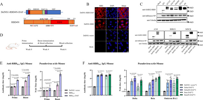
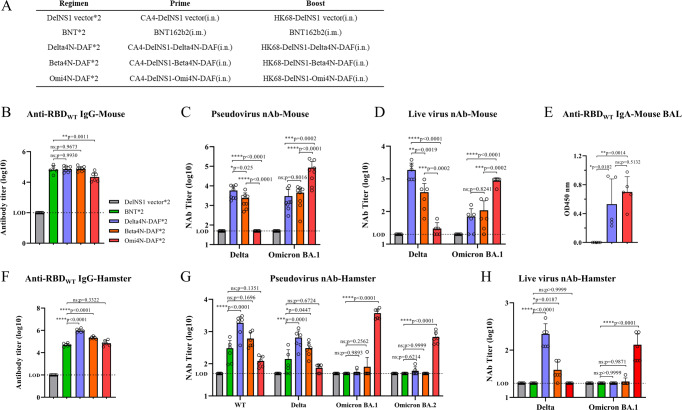
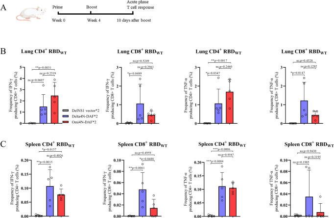
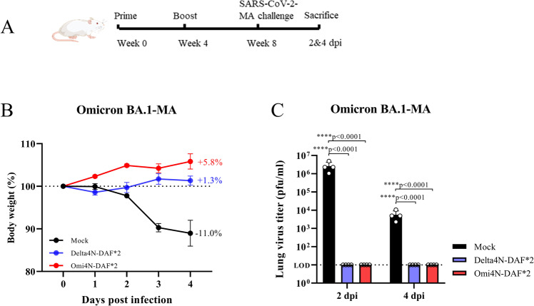
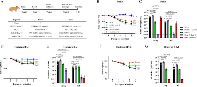
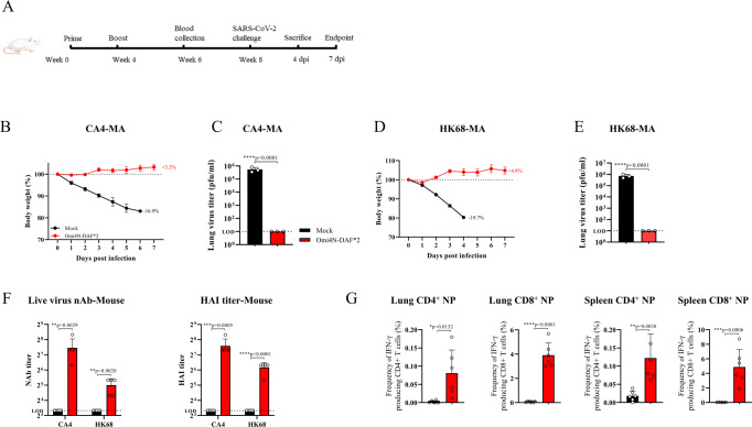
Current available vaccines for COVID-19 are effective in reducing severe diseases and deaths caused by SARS-CoV-2 infection but less optimal in preventing infection. Next-generation vaccines which are able to induce mucosal immunity in the upper respiratory to prevent or reduce infections caused by highly transmissible variants of SARS-CoV-2 are urgently needed. We have developed an intranasal vaccine candidate based on a live attenuated influenza virus (LAIV) with a deleted NS1 gene that encodes cell surface expression of the receptor-binding-domain (RBD) of the SARS-CoV-2 spike protein, designated DelNS1-RBD4N-DAF. Immune responses and protection against virus challenge following intranasal administration of DelNS1-RBD4N-DAF vaccines were analyzed in mice and compared with intramuscular injection of the BioNTech BNT162b2 mRNA vaccine in hamsters. DelNS1-RBD4N-DAF LAIVs induced high levels of neutralizing antibodies against various SARS-CoV-2 variants in mice and hamsters and stimulated robust T cell responses in mice. Notably, vaccination with DelNS1-RBD4N-DAF LAIVs, but not BNT162b2 mRNA, prevented replication of SARS-CoV-2 variants, including Delta and Omicron BA.2, in the respiratory tissues of animals. The DelNS1-RBD4N-DAF LAIV system warrants further evaluation in humans for the control of SARS-CoV-2 transmission and, more significantly, for creating dual function vaccines against both influenza and COVID-19 for use in annual vaccination strategies.


Conflict of interest statement

The authors declare that the University of Hong Kong has filed patents on work related to the generation and application of DelNS1 live attenuated influenza vaccines and the associated platform, with H.C., P.W., and K-Y.Y. included as co-inventors. There is no restriction on the publication of data. The other authors declare that they have no competing interests.

Figures

Fig. 1
Fig. 1. Construction and evaluation of DelNS1-RBD4N-DAF live attenuated virus vaccine candidates.
A Illustration of construction of DelNS1-RBD4N-DAF. B Immunofluorescence (IF) assay of RBD expression with or without DAF in MDCK cells. MDCK cells were mock infected or infected with DelNS1, DelNS1-RBD or DelNS1-RBD-DAF viruses in the backbone of A/California/4/2009 (H1N1) at an MOI of 1 for 10 h and cells processed for IFA using antibodies specific for RBD (red). Nuclei were stained with DAPI (blue). Images are representative of three independent experiments. C Confirmation of N-glycosylation in RBD of DelNS1-RBD4N-DAF LAIV. MDCK cells were mock infected or infected with DelNS1-RBD (WT, lineage A virus), DelNS1-RBD with individual N-glycosylation mutations (A372T, G413N, D428N, or P521N), or DelNS1-RBD4N (4N) LAIV virus at an MOI of 1 for 10 h. Cell lysates were analyzed by western blot using anti-RBD antibody and anti-NP antibody. DelNS1-RBD, DelNS1-RBD4N, and DelNS1 LAIVs were also used to infect A549, BHK21 and MDCK cells to detect the expression of RBD. Images are representative of three independent experiments. D Schedule of immunization and blood collection for BALB/c mice. E Estimation of anti-RBD antibodies in mice prime-boost immunized intranasally with 2 × 10⁶ pfu of DelNS1-RBD-DAF with wild-type (WT) SARS-CoV-2 strain (lineage A) RBD (RBDWT-DAF, n = 4) or DelNS1-RBD with WT-RBD (RBDWT, n = 5) or DelNS1 vector (n = 5). At weeks 4 and week 6, blood was collected from mice and tested for anti-S1 RBD-specific IgG titers by ELISA assay and neutralization activity against pseudovirus expressing wild-type SARS-CoV-2 spike protein. F Estimation of anti-RBD antibodies in mice prime-boost immunized intranasally with 2 × 10⁶ pfu of DelNS1-RBD-DAF with Delta (B.1.617.2) RBD (Delta-DAF, n = 8) or Beta (B.1.351) RBD (Beta-DAF, n = 8), or DelNS1-RBD4N-DAF with Delta RBD4N (Delta4N-DAF, n = 8) or Beta RBD4N (Beta4N-DAF, n = 8), or DelNS1 vector (n = 8). At week 6, blood was collected from mice and tested for anti-S1 RBD-specific IgG titers and neutralization activity against pseudoviruses expressing spike proteins of SARS-CoV-2 variants. LOD: lower limit of detection. Error bars represent mean ± SD. Statistical analysis was performed using one-way ANOVA followed by Dunn’s multiple comparisons test: ****p < 0.0001, **p < 0.01, *p < 0.05, ns not significant. Numerical labels indicate fold difference. Mouse cartoon created with BioRender.com.
Fig. 2
Fig. 2. Immunogenicity of DelNS1-RBD4N-DAF LAIVs in mice and hamsters.
A Prime-boost immunization regimen and grouping of BALB/c mice and hamsters. BALB/c mice were prime-boost immunized intranasally with 2 × 10⁶ pfu of Delta4N-DAF, Beta4N-DAF, DelNS1-RBD4N-DAF with Omicron BA.1 RBD (Omi4N-DAF) or DelNS1 vector only control, or through intramuscular injections of the BioNTech BNT162b2 mRNA vaccine (BNT, 5ugand sera collected 14 days after the second immunization for testing of anti-S1 RBD-specific IgG titers (B) (Delta4N-DAF (n = 8 mice), Beta4N-DAF (n = 8 mice), Omi4N-DAF (n = 6 mice), DelNS1 vector (n = 8 mice), BNT (n = 5 mice)). C Neutralization titers against pseudotyped viruses displaying Delta or Omicron BA.1 spike proteins (n = 8 mice for each group), and (D) neutralization titers against live SARS-CoV-2 variants (Delta and Omicron BA.1) (n = 6 mice for each group). E The bronchoalveolar lavage (BAL) of mice was collected 10 days after boost immunization and anti-S1 RBD-specific IgA titers determined (n = 5 mice for each group). Syrian hamsters were prime-boost immunized intranasally with either 5 × 106 pfu of Delta4N-DAF, Beta4N-DAF, Omi4N-DAF, or DelNS1 vector only control, or through intramuscular injections of the BioNTech BNT162b2 mRNA vaccine (5ug). F Sera samples were collected 14 days after the second immunization and tested for anti-S1 RBD-specific IgG titers (n = 6 hamsters for each group). G Neutralization titers against pseudotyped viruses with wild type, Delta or Omicron spike proteins (n = 6 hamsters for each group), and (H) neutralization titers against live SARS-CoV-2 variants (Delta and Omicron) (n = 6 hamsters for each group). LOD lower limit of detection. Error bars represent mean ± SD. Numerical labels indicate means. Statistical analysis was performed using one-way ANOVA followed by Dunn’s multiple comparisons test: ****p < 0.0001, ***p < 0.001, **p < 0.01, *p < 0.05, ns not significant.
Fig. 3
Fig. 3. DelNS1-RBD4N-DAF LAIV induces CD4+ and CD8+ T cell response.
A Schedule for immunization of BALB/c mice. At day 10 after the second immunization, lung cells and splenocytes were obtained and stimulated with or without spike peptide pools (Supplementary Table 2) overnight in the presence of BFA. Surface markers (CD4, CD8, and Zombie) were stained, and cells were fixed and permeabilized. Intracellular cytokines (TNFα and IFNγ) were then stained with specific antibodies. Delta4N-DAF and Omi4N-DAF induced IFN-γ and TNF-α positive CD4+ and CD8+ T cell responses in lungs (B) and spleens (C) of BALB/c mice. Percentages of IFNγ+ or TNFα+ CD4+ and CD8+ T cells in immunized mice (n = 5 for each group) were compared. Error bars represent mean ± SD. Statistical analysis was performed using one-way ANOVA followed by Dunn’s multiple comparisons test: ***p < 0.001, **p < 0.01, *p < 0.05, ns not significant. Mouse cartoons created with BioRender.com.
Fig. 4
Fig. 4. DelNS1-RBD4N-DAF LAIVs protect mice challenged with mouse-adapted Omicron SARS-CoV-2 variant.
A Illustration of schedule of immunization, SARS-CoV-2 virus challenge and sacrifice for BALB/c mice. 6–8-week-old BALB/c mice were intranasally prime-boost vaccinated with Delta4N-DAF (2 × 10⁶ pfu) or Omi4N-DAF (2 × 10⁶ pfu) or PBS (n = 8 for each group) and then challenged with a mouse-adapted Omicron BA.1 SARS-CoV-2 strain (Omicron-MA, 1 × 105 pfu) 4 weeks after boost immunization. B Body weight changes following virus challenge (n = 8 for each group). C Virus titers in the lungs were measured at 2 dpi (n = 4 for each group) and 4 dpi (n = 4 for each group). MA: mouse-adapted. LOD: lower limit of detection. Error bars represent mean ± SD. Statistical analysis was performed using one-way ANOVA followed by Dunn’s multiple comparisons test: ****p < 0.0001. Mouse cartoon created with BioRender.com.
Fig. 5
Fig. 5. Protection against SARS-CoV-2 virus challenge in hamsters through prime-boost immunization with BNT162b2 mRNA or DelNS1-RBD4N-DAF vaccines.
A Illustration of schedule of immunization, blood collection and SARS-CoV-2 virus challenge for hamsters. Hamsters were prime-boost vaccinated either intranasally with Delta4N-DAF (5 × 106 pfu), Omi4N-DAF (5 × 106 pfu), DelNS1 vector (5 × 106 pfu) or PBS (mock) or intramuscularly with BNT162b2 mRNA (1/6 clinical dose (5ug)) vaccine. Hamsters were challenged with SARS-CoV-2 variants Delta or Omicron BA.2 at 1 × 104 pfu per hamster, 4 weeks after boost immunization. Body weight changes following SARS-CoV-2 virus challenge of hamsters immunized with Delta4N-DAF, Omi4N-DAF or BNT162b2 mRNA vaccines or controls (n = 4 for each group) (B, D, and F). Virus titers in the lungs and nasal turbinates (NT) of hamsters were measured at 4 dpi (n = 4 for each group) (C, E, and G). NT nasal turbinates. LOD lower limit of detection. Error bars represent mean ± SD. Statistical analysis was performed using one-way ANOVA followed by Dunn’s multiple comparisons test: ****p < 0.0001, *p < 0.05, ns not significant. Hamster cartoon created with BioRender.com.
Fig. 6
Fig. 6. Protection against influenza virus challenge in mice prime-boost immunized with DelNS1-RBD4N-DAF LAIV.
A Illustration of schedule of immunization, blood collection and influenza virus challenge for BALB/c mice. BALB/c mice were intranasally prime-boost vaccinated with Omi4N-DAF (2 × 10⁶ pfu) or PBS (n = 6 for each group), and then challenged with mouse-adapted influenza virus strains matching the influenza subtype of vaccines (CA4-) (H1N1, 5 × 103 pfu) or HK68-MA (H3N2, 1 × 104 pfu) (n = 3 for each group)) 4 weeks after boost immunization. Body weight changes in mice following influenza virus challenge were tracked (n = 6 for each group) until the control group reached the body weight loss cut-off (20%) for euthanization, in accordance with animal ethics protocols (B, D). Virus titers in the lungs were measured at 4 dpi (C, E). F BALB/c mice were prime-boost immunized intranasally with 2 × 106 pfu of Omi4N-DAF or PBS (mock) (n = 5 for each group). Sera were collected 14 days after the second immunization for testing of neutralization titers and hemagglutination inhibition (HAI) titers against live influenza viruses CA4 (H1N1) or HK68 (H3N2). G At 5 weeks after the second immunization, 2 μg of PerCP-Cy5.5 conjugated CD45-specific antibody was injected i.v. via the tail vein 5 min before sacrifice. Lung cells and splenocytes were obtained and stimulated with or without influenza NP peptides, overnight in the presence of BFA. Surface markers (CD69, CD103, CD4, CD8, and Zombie) were stained, and cells then fixed and permeabilized. Intracellular IFNγ was then stained with specific antibodies. Omi4N-DAF induced NP-specific tissue-resident memory T (Trm) cell responses in lungs (CD45- IFN-γ+ CD69+ CD4+ T cells and CD45- IFN-γ+ CD69+ CD103+ CD8+ T cells) and spleens (CD45- IFN-γ+ CD4+ and CD8+ T cells). Percentages of T cell subsets in immunized (n = 6) and mock (n = 6) groups were compared. MA mouse-adapted. HAI hemagglutination inhibition. LOD lower limit of detection. Error bars represent mean ± SD. Statistical comparisons between means were performed by Student’s t test (2-tailed): ****p < 0.0001, ***p < 0.001, **p < 0.01, *p < 0.05. Mouse cartoons created with BioRender.com.

Similar articles

Cited by

References

    1. Zhou P, et al. A pneumonia outbreak associated with a new coronavirus of probable bat origin. Nature. 2020;579:270–273. doi: 10.1038/s41586-020-2012-7. - DOI - PMC - PubMed
    1. Taylor L. Covid-19: True global death toll from pandemic is almost 15 million, says WHO. BMJ. 2022;377:o1144. doi: 10.1136/bmj.o1144. - DOI - PubMed
    1. Altmann DM, Boyton RJ. COVID-19 vaccination: the road ahead. Science. 2022;375:1127–1132. doi: 10.1126/science.abn1755. - DOI - PubMed
    1. Subbarao K. The success of SARS-CoV-2 vaccines and challenges ahead. Cell Host Microbe. 2021;29:1111–1123. doi: 10.1016/j.chom.2021.06.016. - DOI - PMC - PubMed
    1. Pulliam JRC, et al. Increased risk of SARS-CoV-2 reinfection associated with emergence of Omicron in South Africa. Science. 2022;376:eabn4947. doi: 10.1126/science.abn4947. - DOI - PMC - PubMed

Publication types

Supplementary concepts