Review
. 2023 May;23(5):295-316.
doi: 10.1038/s41568-023-00560-y. Epub 2023 Apr 12.

Dynamics and specificities of T cells in cancer immunotherapy

Affiliations
Review

Dynamics and specificities of T cells in cancer immunotherapy

Giacomo Oliveira et al. Nat Rev Cancer. 2023 May.

Abstract

Recent advances in cancer immunotherapy - ranging from immune-checkpoint blockade therapy to adoptive cellular therapy and vaccines - have revolutionized cancer treatment paradigms, yet the variability in clinical responses to these agents has motivated intense interest in understanding how the T cell landscape evolves with respect to response to immune intervention. Over the past decade, the advent of multidimensional single-cell technologies has provided the unprecedented ability to dissect the constellation of cell states of lymphocytes within a tumour microenvironment. In particular, the rapidly expanding capacity to definitively link intratumoural phenotypes with the antigen specificity of T cells provided by T cell receptors (TCRs) has now made it possible to focus on investigating the properties of T cells with tumour-specific reactivity. Moreover, the assessment of TCR clonality has enabled a molecular approach to track the trajectories, clonal dynamics and phenotypic changes of antitumour T cells over the course of immunotherapeutic intervention. Here, we review the current knowledge on the cellular states and antigen specificities of antitumour T cells and examine how fine characterization of T cell dynamics in patients has provided meaningful insights into the mechanisms underlying effective cancer immunotherapy. We highlight those T cell subsets associated with productive T cell responses and discuss how diverse immunotherapies might leverage the pre-existing tumour-reactive T cell pool or instruct de novo generation of antitumour specificities. Future studies aimed at elucidating the factors associated with the elicitation of productive antitumour T cell immunity are anticipated to instruct the design of more efficacious treatment strategies.


Conflict of interest statement

Competing interests C.J.W. is an equity holder of BioNTech. G.O. declares no competing interests.

Figures

Fig. 1 |
Fig. 1 |. Phenotypes of CD8+ T cells within the native tumour microenvironment.
Upon activation and recognition of cognate antigens (denoted by the arrow at the top), naive T cells differentiate into antigen-experienced T cells that can be captured by single-cell RNA sequencing (scRNA-seq) as a spectrum of cellular states. As a synthesis of the current literature, we propose a streamlined division of the tumour-infiltrating lymphocyte (TIL) phenotypes into two major compartments: memory T cells (TMem cells; left, blue) and exhausted T cells (TEx cells; right, red, orange and yellow). Of note, such representation does not stringently reflect differentiation pathways and T cell hierarchies, which cannot be fully addressed in human tumour studies due to the lack of longitudinal tracing of T cell clones. TMem cells encompass subsets traditionally subdivided in peripheral blood as stem cell memory T (TSCM), central memory T (TCM) or effector memory T (TEM) cells that maintain signatures of lowly differentiated cells and retain the ability to regenerate a vast progeny of effector cells. In the native tumour microenvironment (TME), TMem cells do not exhibit tumour-specific localization, and this compartment has reduced overall clonal expansion within the TME. Screening of the T cell receptor (TCR) specificity of TMem cells has revealed them to be the preferential reservoir of T cells with antiviral reactivity and, therefore, consistent with bystander TILs, persisting in conditions where the cognate antigen levels are low and controlled. In contrast, TEx cells are highly enriched within the TME, where they demonstrate broad expression of signatures of activation and exhaustion. Their high expression of immune-checkpoint molecules renders them highly sensitive to cancer-induced inhibition. They have been found to consistently express common markers (for example, programmed cell death protein 1 (PD1) and CXC-chemokine ligand 13 (CXCL13)) across studies but display a phenotypic diversity, which translates into a range of capacities for cytotoxicity and extent of dysfunction. Progenitor exhausted T cells (TPE cells) are early dysfunctional T cells that retain the capacity to regenerate a proportion of the TEx cell compartment but lack effector functions. In contrast, terminally exhausted T cells (TTE cells), represent the last stage of differentiation: their cytotoxic capabilities are restrained by inhibitory signals from within the TME due to their high level of expression of immune-checkpoint molecules. Finally, a proportion of TEx cells exhibits expression of signatures related to the acquisition of a tissue-resident memory programme (TRM cells), which confers concomitant expression of inhibitory molecules and high cytotoxic potential. We define this compartment as TRM-like TEx cells because of their phenotypic connections with the TEx cell compartment, but we acknowledge that TRM cells with a similar profile can differentiate in normal tissues upon acute infections to provide a first line of defence against pathogen recurrence. Tumour-specific TRM-like TEx cells have been detected within the TME, thus demonstrating that such a phenotype can also be elicited upon chronic stimulation provided by tumour antigens. Across tumours, the TEx cell compartment is highly clonotypically expanded, consistent with likely recognition of tumour cells. Phenotypic analyses of TCR clonotype families have confirmed the clonal relatedness of diverse TEx cell subsets, possibly constituting different stages of differentiation driven by the recognition of tumour antigens. TEx and TMem TCR clonotypes have been found to only marginally overlap, indicating a distinct division of the specificities of the two compartments. Indeed, several studies of TIL specificity across different tumour types have now unambiguously demonstrated that the TEx cell compartment is almost exclusively enriched in antitumour TCRs. CCR7, CC-chemokine receptor 7; HOBIT, homologue of BLIMP1 in T cell; IL-7R, IL-7 receptor; TCF1, T cell-specific transcription factor 1; TIM3, T cell immunoglobulin mucin receptor 3; TOX, thymocyte selection-associated high mobility group box protein.
Fig. 2 |
Fig. 2 |. Effect of immunotherapies on the T cell repertoire of patients.
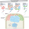
Immunotherapies can rely on antitumour T cells already present within the native tumour microenvironment (TME) or in extratumoural sites (lymph nodes, peripheral blood and normal tissues). Such pre-existing T cell responses (red T cells) can be amplified (indicated by a circular arrow) in extratumoural sites or reinvigorated within the TME (as denoted by the release of cytokines and chemokines) in vivo, through the administration of cancer vaccines or immune-checkpoint blockade (ICB) therapies. Amplification of responses and release of cytokines within the tumour can favour the recruitment of T cells from extratumoural sites. Pre-existing responses can be expanded ex vivo for the generation of T cell products for adoptive tumour-infiltrating lymphocyte (TIL) therapy. By targeting pre-existing responses, which are usually sustained by exhausted T cells specific for a wide array of tumour antigens, such immunotherapies are expected to stimulate polyclonal but dysfunctional T cells. Conversely, the elicitation of non-exhausted antitumour responses requires the de novo generation of antitumour T cells (green T cells). Immunotherapies can elicit de novo T cell responses directly in vivo, through the administration of cancer vaccines that can induce antigen-presenting cell-mediated priming of antigen-unexperienced T cells; alternatively, the specificity of functional T cells may be rewired in vitro by inserting natural T cell receptors (TCRs) or chimeric antigen receptors (CARs) using gene-manipulation technologies. Immunotherapies that induce de novo responses allow the steering of T cell responses towards the recognition of the targeted tumour antigens, optimally under conditions that minimize the induction of exhaustion. Finally, effective antitumour T cells induced by immunotherapies can lyse tumour cells, thus fostering the release of additional tumour antigens, which can be presented by antigen-presenting cells. This phenomenon, known as epitope spreading, can further contribute to the amplification and reinvigoration of pre-existing responses as well as to the induction of new T cell specificities. MHC, major histocompatibility complex.
Fig. 3 |
Fig. 3 |. Mechanisms of response to ICB and the T cell subsets involved.
a, Schema of the characteristics of the native T cell compartment associated with subsequent response (right) or non-response (left) to treatment with immune-checkpoint blockade (ICB). Complete or partial responses to ICB have been associated with a diverse T cell composition within the native tumour microenvironment (TME) as well as with a broader T cell repertoire in extratumoural sites (peripheral blood, normal tissues and draining lymph nodes). Depending on the tumour type, time of analysis and clinical setting, responders can be characterized by high frequencies of intratumoural tissue-resident memory T (TRM) cell-like exhausted T cells with high cytotoxic capacity,,,, increased fractions of progenitor exhausted T (TPE) cells, which possess high regenerative capacity,,, or increased expansion and infiltration of putative tumour-reactive T cells in extratumoural sites. These features are thought to underlie the mechanistic basis of responses to ICB. In contrast, lack of such T cell populations and, consequently, high frequencies of terminally exhausted T (TTE) cells that cannot be substantially reinvigorated predispose patients to ineffective immunotherapy responses. b, Mechanisms of action and dynamics of T cells upon response to ICB in patients. First, by disrupting the inhibitory axis, ICB antibodies can unleash the cytotoxic activity of antitumour TRM cell-like exhausted T cells, which can further expand within the TME (denoted by the circular arrow). This mechanism, so-called ‘cytotoxic revival’, has been observed early after neoadjuvant ICB treatment (2–4 weeks) in head and neck squamous cell carcinoma and breast cancers,,. Second, ICB can have a systemic effect, favouring the intratumoural infiltration of novel (or previously undetected) T cell specificities from extratumoural sites. Newly recruited T cell clones might mount an antitumour response in the presence of ICB antibodies, which could likely prevent the inhibition of cytotoxicity. Such ‘clonal replacement’ was first described as present at a median time of 9 weeks after treatment in patients with basal and squamous cell carcinoma. These systemic dynamics of the T cell repertoire have been observed as predictive of response early after treatment (2–4 weeks) in several clinical settings and cancer types. Finally, response could be sustained by progenitor or early dysfunctional tumour-specific T cells (TPE cells) expanding and differentiating within the TME or recruited from lymph nodes and peripheral tissues. This has been observed in responding lung cancer and melanoma lesions, which were characterized by an accumulation of TPE cells late after treatment (>2 months),. This phenomenon has been termed ‘clonal revival’, since it is characterized by the accumulation of less dysfunctional cells that can potentially regenerate a progeny of effector cells (dashed arrow) capable of tumour cytotoxicity in the presence of the ICB-mediated disruption of inhibitory signals. We propose that such mechanisms might have different kinetics (as presented in the graph modelling the temporal dynamics of T cell responses) and might synergistically contribute to the response to ICB based on the qualitative and quantitative status of the antitumour T cell repertoire (part a).
Fig. 4 |
Fig. 4 |. The phenotypic composition of transferred T cells affects the outcome of adoptive T cell therapy.
The quality of infused T cells with antitumour potential is a major factor contributing to their in vivo persistence and functionality upon transfer to patients with cancer,,,,. Antitumour T cells can be expanded from tumour-infiltrating lymphocytes (TILs) or peripheral blood mononuclear cells (PBMCs), and they can be further manipulated through the introduction of natural T cell receptors (TCRs) or chimeric antigen receptors (CARs) to redirect their specificity towards tumour antigens (left). Ex vivo activation of such T cells results in expansion of T cells with a diverse spectrum of phenotypes (middle), which can affect the dynamics and functionality of T cells once they are transferred into patients with cancer (right). Infusion products enriched in exhausted T (TEx) cell phenotypes with low regenerative potential (terminally exhausted T (TTE) cells and tissue-resident T (TRM) cell-like TEx cells, top right) can provide only short-term antitumour function and rapidly decline in vivo, thus providing a limited control of cancer cells. Indeed, infused TEx cell clones have been shown to contract early following infusion (2–4 weeks) in patients treated with adoptive T cell therapy (ACT) approaches, and the resulting low persistence of the antitumour T cells has been associated with disease progression or recurrence. Conversely, T cell products enriched with memory T (TMem) cells have been associated with high levels of persistence of transferred antitumour T cells and, in turn, with response to ACT (bottom right). The better fitness of infused T cells derives from the presence of cells with regenerative potential (TMem cells) or, to a limited extent, of progenitor exhausted T (TPE) cells, which can expand in vivo, persist long term and differentiate to generate a large number of effectors that provides successful tumour control (dashed arrow). Therefore, ACT can benefit from approaches that favour the ex vivo generation and expansion of non-exhausted antitumour T cells.

References

    1. Sharma P & Allison JP Dissecting the mechanisms of immune checkpoint therapy. Nat. Rev. Immunol 20, 75–76 (2020). - PubMed
    1. Restifo NP, Dudley ME & Rosenberg SA Adoptive immunotherapy for cancer: harnessing the T cell response. Nat. Rev. Immunol 12, 269–281 (2012). - PMC - PubMed
    1. Alcover A, Alarcón B & Di Bartolo V Cell biology of T cell receptor expression and regulation. Annu. Rev. Immunol 36, 103–125 (2018). - PubMed
    1. Kaech SM & Ahmed R Memory CD8+ T cell differentiation: initial antigen encounter triggers a developmental program in naïve cells. Nat. Immunol 2, 415–422 (2001). - PMC - PubMed
    1. Boon T, Coulie PG, Van den Eynde BJ & van der Bruggen P Human T cell responses against melanoma. Annu. Rev. Immunol 24, 175–208 (2006). - PubMed

Publication types