. 2023 Apr;616(7957):534-542.
doi: 10.1038/s41586-023-05729-x. Epub 2023 Apr 12.

The evolution of non-small cell lung cancer metastases in TRACERx

Maise Al Bakir #  1   2 Ariana Huebner #  1   2   3 Carlos Martínez-Ruiz #  1   3 Kristiana Grigoriadis #  1   2   3 Thomas B K Watkins #  2 Oriol Pich #  2 David A Moore  1   2   4 Selvaraju Veeriah  1 Sophia Ward  1   2   5 Joanne Laycock  1 Diana Johnson  1 Andrew Rowan  2 Maryam Razaq  1 Mita Akther  1 Cristina Naceur-Lombardelli  1 Paulina Prymas  1 Antonia Toncheva  1 Sonya Hessey  1   6   7 Michelle Dietzen  1   2   3 Emma Colliver  2 Alexander M Frankell  1   2 Abigail Bunkum  1   6   7 Emilia L Lim  1   2 Takahiro Karasaki  1   2   6 Christopher Abbosh  1 Crispin T Hiley  1   2 Mark S Hill  2 Daniel E Cook  2 Gareth A Wilson  2 Roberto Salgado  8   9 Emma Nye  10 Richard Kevin Stone  10 Dean A Fennell  11   12 Gillian Price  13   14 Keith M Kerr  14   15 Babu Naidu  16 Gary Middleton  17   18 Yvonne Summers  19 Colin R Lindsay  19 Fiona H Blackhall  19 Judith Cave  20 Kevin G Blyth  21   22   23 Arjun Nair  24   25 Asia Ahmed  24 Magali N Taylor  24 Alexander James Procter  24 Mary Falzon  4 David Lawrence  26 Neal Navani  27   28 Ricky M Thakrar  27   28 Sam M Janes  27 Dionysis Papadatos-Pastos  29 Martin D Forster  1   29 Siow Ming Lee  1   29 Tanya Ahmad  29 Sergio A Quezada  1   30 Karl S Peggs  31   32 Peter Van Loo  33   34   35 Caroline Dive  36   37 Allan Hackshaw  38 Nicolai J Birkbak  1   2   39   40   41 Simone Zaccaria  1   7 TRACERx ConsortiumMariam Jamal-Hanjani  42   43   44 Nicholas McGranahan  45   46 Charles Swanton  47   48   49
Collaborators, Affiliations

The evolution of non-small cell lung cancer metastases in TRACERx

Maise Al Bakir et al. Nature. 2023 Apr.

Abstract

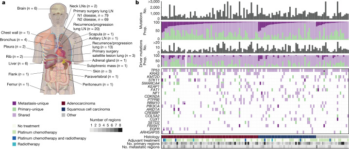
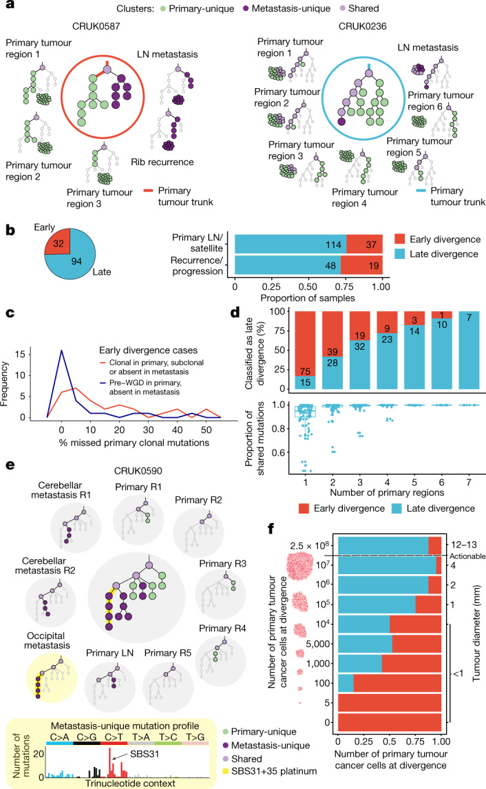
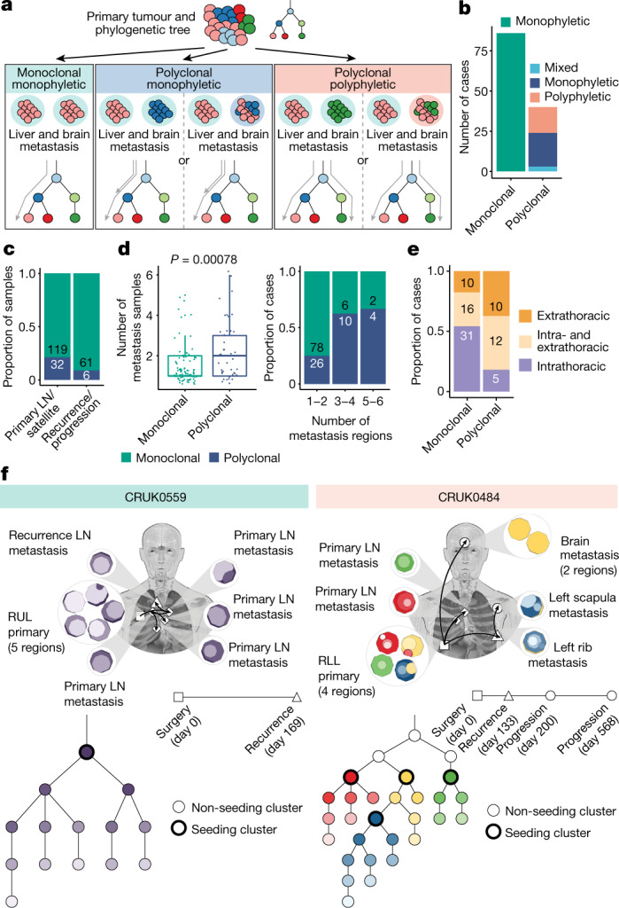
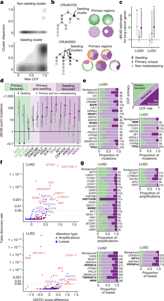
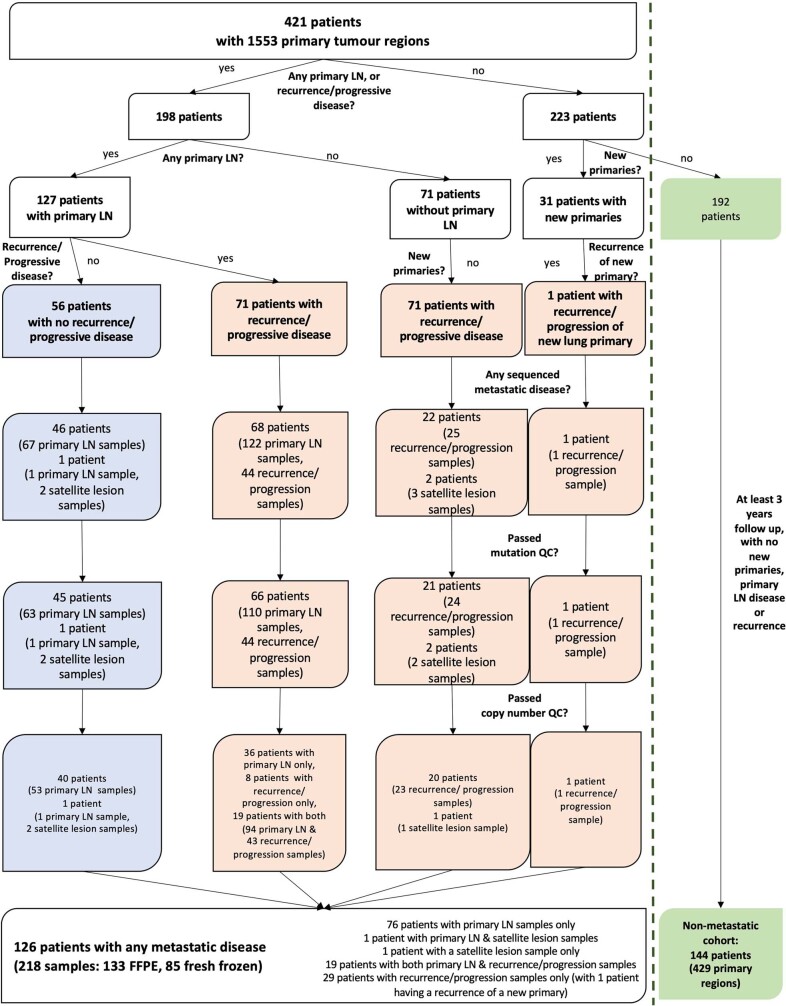
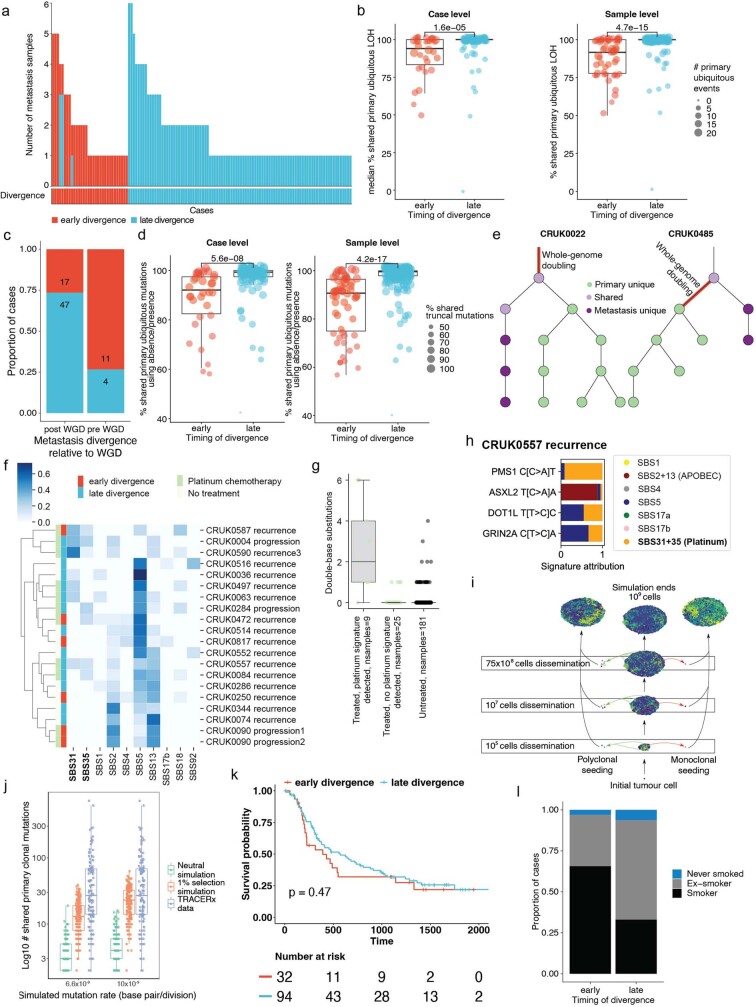
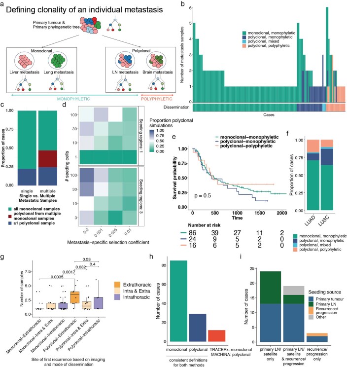
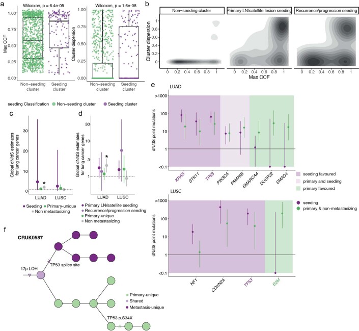
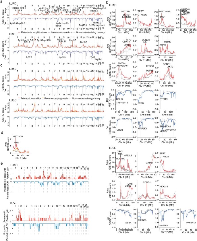
Metastatic disease is responsible for the majority of cancer-related deaths1. We report the longitudinal evolutionary analysis of 126 non-small cell lung cancer (NSCLC) tumours from 421 prospectively recruited patients in TRACERx who developed metastatic disease, compared with a control cohort of 144 non-metastatic tumours. In 25% of cases, metastases diverged early, before the last clonal sweep in the primary tumour, and early divergence was enriched for patients who were smokers at the time of initial diagnosis. Simulations suggested that early metastatic divergence more frequently occurred at smaller tumour diameters (less than 8 mm). Single-region primary tumour sampling resulted in 83% of late divergence cases being misclassified as early, highlighting the importance of extensive primary tumour sampling. Polyclonal dissemination, which was associated with extrathoracic disease recurrence, was found in 32% of cases. Primary lymph node disease contributed to metastatic relapse in less than 20% of cases, representing a hallmark of metastatic potential rather than a route to subsequent recurrences/disease progression. Metastasis-seeding subclones exhibited subclonal expansions within primary tumours, probably reflecting positive selection. Our findings highlight the importance of selection in metastatic clone evolution within untreated primary tumours, the distinction between monoclonal versus polyclonal seeding in dictating site of recurrence, the limitations of current radiological screening approaches for early diverging tumours and the need to develop strategies to target metastasis-seeding subclones before relapse.

Trial registration: ClinicalTrials.gov NCT01888601.


Conflict of interest statement

M.A.B. has consulted for Achilles Therapeutics. D.A.M. reports speaker fees from AstraZeneca, Eli Lilly and Takeda, consultancy fees from AstraZeneca, Thermo Fisher Scientific, Takeda, Amgen, Janssen, MIM Software, Bristol Myers Squibb (BMS) and Eli Lilly and has received educational support from Takeda and Amgen. S.V. is listed as a co-inventor on a patent for detecting molecules in a sample (U.S. patent no. 10578620). A.M.F. is listed as a co-inventor on a patent application to determine methods and systems for tumour monitoring (PCT/EP2022/077987). C.A. has received speaking honoraria or expenses from Novartis, Roche, AstraZeneca and BMS and reports employment at AstraZeneca; is listed as an inventor on a European patent application relating to assay technology to detect tumour recurrence (PCT/GB2017/053289), the patent has been licensed to commercial entities and, under his terms of employment, C.A is due a revenue share of any revenue generated from such license(s); declares a patent application (PCT/US2017/028013) for methods to detect lung cancer; and is a named inventor on a patent application to determine methods and systems for tumour monitoring (PCT/EP2022/077987); and is a named inventor on a provisional patent protection related to a ctDNA detection algorithm. C.T.H. has received speaker fees from AstraZeneca. G.A.W. is employed by and has stock options in Achilles Therapeutics. R.S. reports non-financial support from Merck and BMS, research support from Merck, Puma Biotechnology and Roche, and personal fees from Roche, BMS and Exact Sciences for advisory boards. D.A.F reports grants from Aldeyra, Boehringer Ingelheim, Astex Therapeutics, Bayer, BMS, GSK, RS Oncology, Clovis, Eli Lilly, BMS, MSD and GSK, and personal fees from Atara, BMS, Boehringer Ingelheim, Cambridge Clinical Laboratories, Targovax, Roche and RS Oncology. C.R.L. has provided consulting/advisory support to Amgen and Hanson Wade, and educational support to Amgen; and has received financial support for research from Revolution Medicines, as well as non-financial support from Amgen. J.C. reports funding from Amgen to attend a conference. A.N. reports personal fees from Aidence BV and Faculty Science Limited. N.N. reports honoraria for non-promotional educational talks, advisory boards or conference attendance from Amgen, AstraZeneca, Boehringer Ingelheim, BMS, Fujifilm, Guardant Health, Intuitive, Janssen, Lilly, Merck Sharp & Dohme, Olympus, OncLive, PeerVoice, Pfizer and Takeda. S.M.J. has received fees for advisory board membership in the last three years from Astra-Zeneca, Bard1 Lifescience and Johnson and Johnson, grant income from Owlstone and GRAIL, and assistance with travel to an academic meeting from Cheisi. M.D.F. acknowledges grant support from CRUK, AstraZeneca, Boehringer Ingelheim, MSD and Merck; is an advisory board member for Transgene; and has consulted for Achilles, Amgen, AstraZeneca, Bayer, Boxer, BMS, Celgene, EQRx, Guardant Health, Immutep, Ixogen, Janssen, Merck, MSD, Nanobiotix, Novartis, Oxford VacMedix, Pharmamar, Pfizer, Roche, Takeda and UltraHuman. S.A.Q. is a co-founder, stockholder and chief scientific officer of Achilles Therapeutics. K.S.P is a co-founder of Achilles Therapeutics. C.D. received research funding/educational research grants from AstraZeneca, Astex Pharmaceuticals, Bioven, Amgen, Carrick Therapeutics, Merck, Taiho Oncology, GSK, Bayer, Boehringer Ingelheim, Roche, BMS, Novartis, Celgene, Epigene Therapeutics, Angle, Menarini, Clearbridge Biomedics, Thermo Fisher Scientific and Neomed Therapeutics; and received honoraria for consultancy and/or advisory boards from Biocartis, Merck, AstraZeneca, GRAIL and Boehringer Ingelheim. A.Ha. has received fees for being a member of independent data monitoring committees for Roche-sponsored clinical trials, and academic projects co-ordinated by Roche. N.J.B. is listed as a co-inventor on a patent to identify responders to cancer treatment (PCT/GB2018/051912) and a co-inventor on a patent for methods for predicting anti-cancer response (US14/466,208). M.J.-H. has consulted for, and is a member of, the Achilles Therapeutics scientific advisory board and steering committee, has received speaker honoraria from Pfizer, Astex Pharmaceuticals and Oslo Cancer Cluster, and is listed as a co-inventor on a European patent application relating to methods to detect lung cancer PCT/US2017/028013); this patent has been licensed to commercial entities and, under terms of employment, M.J.-H. is due a share of any revenue generated from such license(s). N.M. has received consultancy fees and has stock options in Achilles Therapeutics; and holds European patents relating to targeting neoantigens (PCT/EP2016/059401), identifying patient response to immune checkpoint blockade (PCT/EP2016/071471), determining HLA LOH (PCT/GB2018/052004), and predicting survival rates of patients with cancer (PCT/GB2020/050221). C.S. acknowledges grant support from AstraZeneca, Boehringer-Ingelheim, BMS, Pfizer, Roche-Ventana, Invitae (previously Archer Dx, collaboration in minimal residual disease sequencing technologies), Ono Pharmaceutical, and Personalis; is an AstraZeneca advisory board member and chief investigator for the AZ MeRmaiD 1 and 2 clinical trials and is also co-chief investigator of the NHS Galleri trial funded by GRAIL and a paid member of GRAIL’s scientific advisory board; receives consultant fees from Achilles Therapeutics (also a scientific advisory board member), Bicycle Therapeutics (also a scientific advisory board member), Genentech, Medicxi, China Innovation Centre of Roche (CICoR) formerly Roche Innovation Centre – Shanghai, Metabomed (until July 2022) and the Sarah Cannon Research Institute; has received honoraria from Amgen, AstraZeneca, Pfizer, Novartis, GlaxoSmithKline, MSD, Bristol Myers Squibb, Illumina, and Roche-Ventana; had stock options in Apogen Biotechnologies and GRAIL until June 2021, and currently has stock options in Epic Bioscience, Bicycle Therapeutics, and has stock options and is co-founder of Achilles Therapeutics; is listed as an inventor on a European patent application relating to assay technology to detect tumour recurrence (PCT/GB2017/053289), the patent has been licensed to commercial entities and, under his terms of employment, C.S. is due a revenue share of any revenue generated from such license(s); holds patents relating to targeting neoantigens (PCT/EP2016/059401), identifying patient response to immune checkpoint blockade (PCT/EP2016/071471), determining HLA LOH (PCT/GB2018/052004), predicting survival rates of patients with cancer (PCT/GB2020/050221), identifying patients who respond to cancer treatment (PCT/GB2018/051912), a US patent relating to detecting tumour mutations (PCT/US2017/28013), methods for lung cancer detection (US20190106751A1) and both a European and US patent related to identifying insertion/deletion mutation targets (PCT/GB2018/051892) and is listed as a co-inventor on a patent application to determine methods and systems for tumour monitoring (PCT/EP2022/077987) and is a named inventor on a provisional patent protection related to a ctDNA detection algorithm.

Figures

Fig. 1
Fig. 1. Sample distribution and mutational overview in the paired primary metastasis TRACERx 421 cohort.
a, The distribution of metastatic samples by anatomical location; n indicates number of samples used in analyses. b, The total number of mutations and putative driver mutations detected per case (grey bars) and the proportion of these mutations that are unique to the primary tumour (green) or metastasis (dark purple), or shared between primary and metastasis (light purple) per case. The top 20 most frequently mutated cancer genes and their presence/absence in the primary and metastatic samples, including instances of two driver mutations in the same gene, are also shown. The histology, number of primary and metastatic samples sequenced, and adjuvant therapy status is illustrated. No., number; prop., proportion; LN, lymph node.
Fig. 2
Fig. 2. Timing metastatic divergence.
a, Example phylogenetic trees depicting early (CRUK0587) and late (CRUK0236) divergence. Light purple, shared; dark purple, metastasis-unique; green, primary-unique mutation clusters. b, Pie chart showing the fraction of cases with early (= 32) and late divergence (n = 94) (left panel). The proportion of early and late divergence by metastasis type at the sample level (primary LN/satellite versus recurrence/progression; Fisher’s exact test, P = 0.61)  (right panel). c, In early divergence cases, the median number of pre-WGD mutations (blue line) defined as not clonal in the metastases is 1.4% (IQR = 0.8–3.2%; n = 28). Post-WGD mutations or mutations in non-WGD tumours (red line) were more likely to be not clonal in the metastasis (median = 8.5%, IQR = 3.0–22.3%; n = 32; Wilcoxon rank-sum test, P = 0.003). d, Downsampling of late divergence cases (n = 94). A random set of primary tumour regions was used to re-classify the timing of divergence for each case (top panel); proportion of shared mutations between downsampled primary tumour and metastases (bottom panel). e, Phylogenetic trees for case CRUK0590 (inner circle) and within each region, depicting an active platinum signature in the occipital metastasis, timing metastatic divergence of the occipital and cerebellar metastases to a period when platinum therapy was delivered, approximately 6–8 months before recurrence. f, Simulations of tumour size (n = 20 simulations per tumour size) at metastatic clone divergence suggest that early divergence is more likely to happen when the primary tumour is small; a diameter ≥8 mm is a typical threshold used to investigate solid nodules detected using computed tomography (denoted ‘actionable’). The box plots represent the upper and lower quartiles (box limits), the median (centre line) and the vertical bars span the 5th to 95th percentiles. All tests were two-sided unless otherwise specified. R, region; LN, lymph node; WGD, whole genome doubling.
Fig. 3
Fig. 3. Modes of dissemination.
a, Definitions of the dissemination patterns of metastases at the case level, described relative to the primary tumour phylogeny. Grey arrows indicate the branches leading up to the seeding cluster(s). b, The most prevalent mode of metastatic dissemination observed is monoclonal monophyletic. Polyclonal ‘mixed’ represents cases in which a consensus dissemination pattern could not be inferred due to different possible phylogenetic tree topologies. c, At the sample level, polyclonal dissemination is more prevalent in primary LN/satellite lesions compared to recurrence/progression lesions (Fisher’s exact test, P = 0.03). d, Polyclonal dissemination is associated with a higher number of metastatic samples compared with monoclonal dissemination (median number of metastasis samples: 2 versus 1, respectively; Wilcoxon rank-sum test, P = 0.00078), sample number depicted as discrete values (left panel) or proportion (right panel). e, In cases where recurrence occurs, polyclonal dissemination is associated with extrathoracic metastasis, as identified on imaging, compared with monoclonal dissemination (n = 57 (monoclonal), n = 27 (polyclonal); Fisher’s exact test, P = 0.0056) f, Examples of cases with monoclonal (CRUK0559) and polyclonal polyphyletic (CRUK0484) dissemination patterns, both of which also demonstrate metastases being seeded from other sites of metastatic disease. The black arrows on the body map represent the routes of metastatic seeding (MACHINA). Each seeding cluster in the phylogenetic tree, as defined by our method, is assigned a unique colour that is also represented in the region clone maps. The timeline indicates the day on which the metastases were detected on imaging; the biopsy dates differ from this. For CRUK0559, the recurrence biopsy took place on day 188. For CRUK0484, the rib recurrence, scapula progression and brain progression were sampled at days 147, 433 and 582, respectively. The box plots represent the upper and lower quartiles (box limits), the median (centre line) and the vertical bars span the 5th to 95th percentiles. All tests were two-sided unless otherwise specified. LN, lymph node; RUL, right upper lobe; RLL, right lower lobe.
Fig. 4
Fig. 4. Selection in metastasis.
a, Cluster dispersion and maximum cancer cell fraction (CCF) across primary tumour regions in the subclonal seeding clusters versus non-seeding clusters in metastasizing tumours. b, Examples of seeding cluster dispersion across primary tumour regions illustrated by one clone-map per region. CRUK0702 demonstrates a single dominant seeding cluster (purple), dispersed across two primary tumour regions. CRUK0063 highlights two dominant (purple and yellow) and one minor seeding cluster (pink). c, Cohort-level selection (n = 111 genes) of seeding (purple) versus primary-unique mutations from metastasizing tumours (green) versus subclonal non-metastasizing primary tumour mutations (grey). The dots represent dN/dS estimates; the asterisks indicate values that are significantly different from 1. d, Gene-level dN/dS values of seeding mutations versus combined primary-unique/non-metastasizing primary tumour mutations for all histologies. A dN/dS odds ratio (OR) of >2 indicates a seeding favoured gene; <0.5 is primary favoured; 0.5–2 is classified as both primary and seeding favoured. Purple and green gene names represent significant enrichment in seeding and non-seeding mutations, respectively. The lines indicate the 95% CIs. e, Paired primary tumour–metastasis (met) mutation analysis. Metastasis favoured mutations are defined as having a higher clonality in metastases compared with the primary tumour; primary favoured if the clonality is higher in the primary tumour; the remaining were classified as maintained; background refers to mutations in non-cancer genes. f, The GISTIC2.0 score difference between the unpaired metastases and non-metastasizing cohorts plotted against the false-discovery rate of the G-score in the metastases cohort for cancer genes. Amplified genes are shown in red; deleted genes are shown in blue. Horizontal dotted lines indicate p = 0.05 g, Paired SCNA analysis of cancer genes that were found to be significant in f. An amplification/deletion was classified as metastasis favoured if it was present in the metastasis and absent in the primary tumour, primary favoured if present in the primary tumour but not the metastasis, or otherwise defined as maintained. Only tumours that had at least one copy number event in the gene in any sample were counted. For e and g, significant genes (multinomial test; p < 0.05) are shown in bold; asterisks represent significance after multiple-testing correction (q < 0.05); numbers in parentheses indicate number of events.
Extended Data Fig. 1
Extended Data Fig. 1. Cohort and sample overview.
Sample acquisition and quality control overview, also highlighting the non-metastatic cohort. In addition to the 31 patients with new primary tumours highlighted in the figure, there are 10 other new primary cases within the metastatic cohort, totalling 41 new primary cases; LN, lymph node; QC, quality control; FFPE, formalin-fixed paraffin embedded tissue.
Extended Data Fig. 2
Extended Data Fig. 2. Genomic analyses of primary and metastases.
a. Comparison of primary tumour and metastasis purity (median purity: primary = 0.43, primary LN/satellite = 0.32, recurrence/progression = 0.31, Wilcoxon rank-sum test), b. Comparison of primary tumour and metastasis ploidy (median ploidy: primary = 3.1, primary LN/satellite = 2.95, recurrence/progression = 3.1, Wilcoxon rank-sum test) c. Comparison of primary tumour and metastasis weighted genomic instability index (wGII, median wGII: primary = 0.53, primary LN/satellite = 0.52, recurrence/progression = 0.51, Wilcoxon rank-sum test) d. Comparison of primary tumour and metastasis fraction of the genome subject to loss of heterozygosity (FLOH, median FLOH: primary = 0.28, primary LN/satellite = 0.28, recurrence/progression = 0.32, Wilcoxon rank-sum test) e. Comparison of primary tumour and metastasis tumour mutation burden (TMB, median TMB: primary = 508, primary LN/satellite = 479, recurrence/progression = 651, Wilcoxon rank-sum test). f. Comparison of whole genome doubling (WGD) status between primary tumours and paired metastases. There is no enrichment in WGD in metastases (Fisher’s exact test, p = 1). The box plots represent the upper and lower quartiles (box limits), the median (centre line) and the vertical bars span the 5th to 95th percentiles. All tests were two-sided unless otherwise specified.
Extended Data Fig. 3
Extended Data Fig. 3. Overview of all phylogenetic trees.
All phylogenetic trees for the 126 tumours with their paired metastases, split by timing of divergence (early vs. late). Clusters annotated in green are primary-unique, clusters in light purple are shared, while clusters in dark purple are metastasis-unique.
Extended Data Fig. 4
Extended Data Fig. 4. Timing of metastatic divergence.
a. Sample level divergence timing (early and late). Where both early and late divergence is seen in multiple metastasis samples of one case, the overall timing is defined as early. b. Orthogonal method to time metastatic divergence using primary ubiquitous arm-level loss of heterozygosity (LOH). Arm level LOH was significantly more likely to be fully clonal in late compared to early divergence (case level median early = 0.94, late = 1, Wilcoxon rank-sum test, p = 1.6e-5; sample level median, early = 0.92, late = 1, Wilcoxon rank-sum test, p = 4.7e-15). c. Orthogonal method timing divergence using primary clonal whole genome doubling (WGD). There is enrichment of early divergence in pre-WGD divergence (Fisher’s exact test, p = 0.0017). d. Orthogonal method to time metastatic divergence using simple absence/presence of mutations in the primary tumour, to define primary ubiquitous mutations. Early divergent tumours have a lower proportion of shared primary ubiquitous mutations (case level median early = 92.1%, late = 99.3%, Wilcoxon rank-sum test, p = 5.6e-8; sample level median, early = 90.7%, late = 99.6%, Wilcoxon rank-sum test, p = 4.20e-17). e. Examples of pre- and post-WGD divergence (CRUK0485 and CRUK0022, respectively). The red line represents the branch with WGD. f. Detected mutational signatures using sample unique mutations for each of the metastatic samples with sufficient mutations (more than 50). SBS31 and SBS35 represent the platinum mutation signatures. g. In patients treated with platinum chemotherapy and where platinum signature was detected in the metastases (9 samples), an enrichment was seen in sample-specific double base substitutions (Mann-Whitney-U test; treated and detected platinum signature vs. treated and no signature detected (25 samples), p = 2.58e-5; treated and detected platinum signature vs. untreated (181 samples), p = 1.32e-10). h. In cases where platinum signature was detected, putative metastasis-unique driver mutations were mapped to the most likely signature. Example case of CRUK0557 where mapping such mutations (PMS1, ASXL2, DOT1L, GRIN2A) revealed PMS1 to likely be platinum-driven. i. Schematic representation of the agent-based modelling approach used to investigate timing and patterns of metastatic seeding. j. Number of shared primary clonal mutations between simulated primary-metastasis pairs and the different mutations and selection rates. Additionally, the number of shared primary clonal mutations from TRACERx data is indicated. k. Kaplan-Meier analysis demonstrating no significant difference in early vs. late divergence (Log rank test, p = 0.47). l. Early divergence is associated with a higher proportion of current smokers (n early = 32, n late = 94; Fisher’s exact test, p = 0.005). The box plots represent the upper and lower quartiles (box limits), the median (centre line) and the vertical bars span the 5th to 95th percentiles. All tests were two-sided unless otherwise specified.
Extended Data Fig. 5
Extended Data Fig. 5. Modes of dissemination.
a. Sample level definitions of dissemination patterns relative to the primary tumour phylogeny. b. Sample level dissemination patterns with overall case level defined beneath c. Proportion of cases defined as polyclonal or monoclonal divided by whether a single or multiple metastatic samples were available (n single = 77, n multiple = 49). There is increased power to detect polyclonal seeding when multiple metastatic samples were sequenced (in dark red, we see approximately 22.4% of polyclonal cases result from multiple monoclonal seeding patterns). d. Proportion of observed polyclonal metastases when simulating differing numbers of disseminating primary tumour cells (y-axis) and varying the number of primary regions from which this occurs (top and bottom panel). The primary tumour was always simulated with 1% selection while the selection coefficients were varied in the metastasis (x-axis). Increasing selection pressure in the metastasis is associated with the appearance of monoclonal dissemination even if the dissemination from the primary tumour is polyclonal. The fewer the number of disseminating cells, the stronger the effect. e. Kaplan-Meier analyses demonstrate no significant difference in lung-cancer specific disease-free survival across the different dissemination patterns (Log rank test, p = 0.5). f. Proportion of dissemination type on a case level, as seen in the main histologic subtypes (LUAD, n = 65; LUSC, n = 39; Fisher’s exact test, p > 0.05). g. Tumours with polyclonal dissemination and extrathoracic metastases have more metastatic samples acquired (Wilcoxon rank-sum test). h. Comparison of the TRACERx dissemination definitions with MACHINA shows that the majority of dissemination patterns are consistent across the two methods, with only 12/126 cases differing; with the TRACERx definitions being more conservative by classifying these cases as monoclonal whereas MACHINA defines these as polyclonal. i. Summary of MACHINA analysis of a metastasis seeding other sites of disease in 46 cases with multiple metastatic samples. ‘Other’ represents cases where the primary tumour seeds the recurrence and additional metastasis seeding patterns are concurrently observed (e.g., recurrence/progression sample seeding the primary LN, primary LN to primary LN seeding, recurrence seeding a progression sample). The box plots represent the upper and lower quartiles (box limits), the median (centre line) and the vertical bars span the 5th to 95th percentiles. All tests were two-sided unless otherwise specified; LN, lymph node.
Extended Data Fig. 6
Extended Data Fig. 6. Mutation selection in metastases.
a. Comparison of maximum cancer cell fraction (CCF) in subclonal primary-unique and seeding clusters (Wilcoxon rank-sum test, p = 6.4e-5) and clonal dispersion of primary-unique and seeding clusters (Wilcoxon rank-sum test, p = 1.6e-8). b. Higher dispersion and CCF is seen in the seeding clusters of both primary LN/satellite lesions and recurrence/progression samples compared to non-seeding clusters. Clusters that are found in both primary LN/satellite lesions and recurrence/progression samples were excluded from this analysis. c. Cohort level selection (n genes = 111) of only subclonal mutations in seeding vs. primary-unique mutations vs. mutations in non-metastasizing primary tumours. d. Cohort level selection (n genes = 111) of primary LN/satellite lesions vs. recurrence/progression seeding mutations vs. primary-unique mutations vs. mutations in non-metastasizing primary tumours. Dots represent dN/dS estimates; the asterisks indicate values that are significantly different from 1. e. Gene-level dN/dS values of seeding mutations vs primary-unique and non-metastasizing primary tumour mutations split by lung adenocarcinoma (LUAD) and lung squamous cell carcinoma (LUSC). Genes are classified as seeding favoured if the odds ratio (OR) of dN/dS of seeding vs. primary-unique mutations >2, primary favoured if OR <0.5, and otherwise classified as both primary and seeding favoured. Genes highlighted in purple and green are significantly enriched in seeding and non-seeding mutations respectively. f. Phylogenetic tree of CRUK0587. Clusters annotated in green are primary-unique, clusters in light purple are shared, while clusters in dark purple are metastasis-unique. There is a metastasis-unique TP53 splice site mutation which occurred independently of a primary-unique S34X TP53 mutation. Lines indicate the 95% confidence intervals for c,d and e. The box plots represent the upper and lower quartiles (box limits), the median (centre line) and the vertical bars span the 5th to 95th percentiles. All tests are two-sided unless otherwise specified.
Extended Data Fig. 7
Extended Data Fig. 7. Somatic copy number aberration selection in metastases.
a. Across-genome GISTIC2.0 scores are plotted for amplifications and deletions for lung adenocarcinoma (LUAD) and lung squamous cell carcinoma (LUSC). Annotated cytobands contain genes overlapping loci with significant G-scores in the metastasis cohort and that have a GISTIC2.0 score difference (GSD) >0 between unpaired metastases and non-metastasizing tumours. b. Individual chromosome plots highlighting genes overlapping significant loci in the metastasis cohort with GSD > 0 that were detected in the unpaired analysis performed in a. c. Across-genome GISTIC2.0 scores are plotted for amplifications and deletions for LUAD and LUSC separating primary LN/satellite lesions and recurrence/progression samples. d. Individual plot highlighting GSD between primary LN/satellite lesions, recurrence/progression samples and non-metastatic primary regions on chromosome 6 encompassing HIST1H3B. The locus is significantly amplified in the primary LN/satellite lesions (GSD = 1.90, q = 1.30e-7). e. Across genome plot showing the frequency of parallel gains/amplification events in red, and frequency of parallel loss/LOH events in blue. The top and bottom panels show the parallel evolution between primary regions harbouring the seeding clone and their paired metastases in LUAD and LUSC respectively; Amp, amplification; Del, deletion; Chr, chromosome; Mb, megabase.

Comment in

References

    1. Bray, F. et al. Global cancer statistics 2018: GLOBOCAN estimates of incidence and mortality worldwide for 36 cancers in 185 countries. CA Cancer J. Clin.68, 394–424 (2018). - PubMed
    1. Zappa, C. & Mousa, S. A. Non-small cell lung cancer: current treatment and future advances. Transl. Lung Cancer Res.5, 288–300 (2016). - PMC - PubMed
    1. Jamal-Hanjani, M. et al. Tracking the evolution of non–small-cell lung cancer. N. Engl. J. Med.376, 2109–2121 (2017). - PubMed
    1. Grigoriadis, K. et al. CONIPHER: a computational framework for scalable phylogenetic reconstruction with error correction. Protoc. Exchange10.21203/rs.3.pex-2158/v1 (2023). - PubMed
    1. de Bruin, E. C. et al. Spatial and temporal diversity in genomic instability processes defines lung cancer evolution. Science346, 251–256 (2014). - PMC - PubMed

Publication types

Associated data