

Vitamin D receptor prevents tumour development by regulating the Wnt/β-catenin signalling pathway in human colorectal cancer
- PMID: 37046222
- PMCID: PMC10091620
- DOI: 10.1186/s12885-023-10690-z
Vitamin D receptor prevents tumour development by regulating the Wnt/β-catenin signalling pathway in human colorectal cancer
Abstract
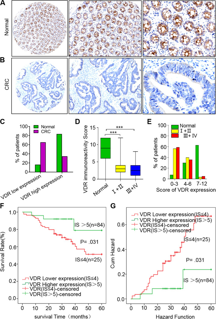
Background: Colorectal cancer (CRC) is a common disease threatening human lives worldwide, and vitamin D receptor (VDR) contributes protective roles in this disease. However, the molecular mechanisms underlying VDR protection in CRC progression require further investigation.
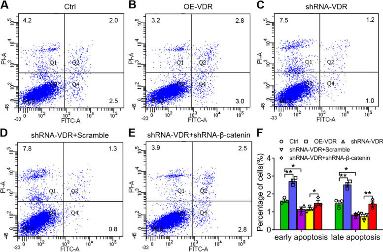
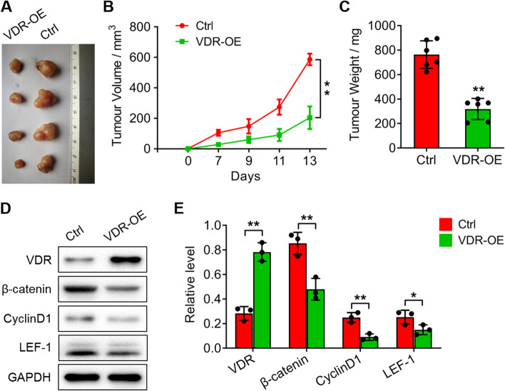
Methods: In this study, we statistically analyzed the relationship between VDR expression and CRC development in patients and detected invasion and apoptosis in CRC cells with VDR overexpression and interference. We also detected the expression of key genes involved in Wnt/β-catenin signalling (β-catenin, lymphoid enhancer factor (LEF)-1 and cyclin D1) in SW480 cells and nude mice injected with VDR-overexpressing SW480 cells and observed tumour development. Additionally, we performed Co-immunoprecipitation (Co-IP) and glutathione-S-transferase (GST) pull-down assays to identify the protein interactions of VDR with β-catenin, dual luciferase (LUC) and chromatin immunoprecipitation (ChIP) to detect the activation of LEF-1 by VDR.
Results: The VDR level was closely related to the development and prognosis of CRC patients. VDR overexpression inhibited invasion but promoted apoptosis in cancer cells. β-catenin shRNA contributed oppositely to cancer cell activity with VDR shRNA. Additionally, VDR interacted with β-catenin at the protein level and blocked its nuclear accumulation. VDR regulated the expression of β-catenin, cyclin D1 and LEF-1 and directly activated LEF-1 transcription in vitro. Furthermore, nude mice injected with VDR-overexpressing SW480 cells revealed suppression of tumour growth and decreased expression of β-catenin, cyclin D1 and LEF-1.
Conclusions: This study indicated that VDR protected against CRC disease in humans by inhibiting Wnt/β-catenin signalling to control cancer cell invasion and apoptosis, providing new evidence to explore VDR biomarkers or agonists for CRC patient diagnosis and treatment.
Keywords: Apoptosis; Colorectal cancer; Cyclin D1; Invasion; Vitamin D receptor; Wnt/β-catenin.
© 2023. The Author(s).
Conflict of interest statement
The authors declare that there are no relevant conflicts of interest.
Figures


Similar articles
-
Histone Demethylase JMJD2D Interacts With β-Catenin to Induce Transcription and Activate Colorectal Cancer Cell Proliferation and Tumor Growth in Mice.Gastroenterology. 2019 Mar;156(4):1112-1126. doi: 10.1053/j.gastro.2018.11.036. Epub 2018 Nov 23. Gastroenterology. 2019. PMID: 30472235
-
SPDEF Induces Quiescence of Colorectal Cancer Cells by Changing the Transcriptional Targets of β-catenin.Gastroenterology. 2017 Jul;153(1):205-218.e8. doi: 10.1053/j.gastro.2017.03.048. Epub 2017 Apr 5. Gastroenterology. 2017. PMID: 28390865 Free PMC article.
-
TRIB3 Interacts With β-Catenin and TCF4 to Increase Stem Cell Features of Colorectal Cancer Stem Cells and Tumorigenesis.Gastroenterology. 2019 Feb;156(3):708-721.e15. doi: 10.1053/j.gastro.2018.10.031. Epub 2018 Oct 24. Gastroenterology. 2019. PMID: 30365932
-
Mechanisms of action of vitamin D in colon cancer.J Steroid Biochem Mol Biol. 2019 Jan;185:1-6. doi: 10.1016/j.jsbmb.2018.07.002. Epub 2018 Jul 4. J Steroid Biochem Mol Biol. 2019. PMID: 29981368 Review.
-
Mechanism of action of vitamin D and the vitamin D receptor in colorectal cancer prevention and treatment.Rev Endocr Metab Disord. 2012 Mar;13(1):31-8. doi: 10.1007/s11154-011-9196-y. Rev Endocr Metab Disord. 2012. PMID: 21861107 Free PMC article. Review.
Cited by
-
Vitamin D improves autoimmune diseases by inhibiting Wnt signaling pathway.Immun Inflamm Dis. 2024 Feb;12(2):e1192. doi: 10.1002/iid3.1192. Immun Inflamm Dis. 2024. PMID: 38414312 Free PMC article.
-
A comprehensive AI-driven analysis of large-scale omic datasets reveals novel dual-purpose targets for the treatment of cancer and aging.Aging Cell. 2023 Dec;22(12):e14017. doi: 10.1111/acel.14017. Epub 2023 Oct 27. Aging Cell. 2023. PMID: 37888486 Free PMC article.
-
Calcitriol Treatment Decreases Cell Migration, Viability and β-Catenin Signaling in Oral Dysplasia.Curr Issues Mol Biol. 2024 Apr 2;46(4):3050-3062. doi: 10.3390/cimb46040191. Curr Issues Mol Biol. 2024. PMID: 38666921 Free PMC article.
-
Unraveling colorectal cancer prevention: The vitamin D - gut flora - immune system nexus.World J Gastrointest Oncol. 2024 Jun 15;16(6):2394-2403. doi: 10.4251/wjgo.v16.i6.2394. World J Gastrointest Oncol. 2024. PMID: 38994172 Free PMC article. Review.
-
Key genes of vitamin D metabolism and their roles in the risk and prognosis of cancer.Front Genet. 2025 Jun 24;16:1598525. doi: 10.3389/fgene.2025.1598525. eCollection 2025. Front Genet. 2025. PMID: 40630116 Free PMC article. Review.
References
-
- Svestka T, Krechler T. Preventing colorectal cancer. Cas Lek Cesk. 2016;155:27–29. - PubMed
-
- Haraldsdottir S, Einarsdottir HM, Smaradottir A, Gunnlaugsson A, Halfdanarson TR. [Colorectal cancer - review]. Laeknabladid. 2014; 100: 75–82. - PubMed
-
- Zheng Y, Trivedi T, Lin RC, Fong-Yee C, Nolte R, Manibo J, Chen Y, Hossain M, Horas K, Dunstan C, et al. Loss of the vitamin D receptor in human breast and prostate cancers strongly induces cell apoptosis through downregulation of Wnt/β-catenin signaling. Bone Res. 2017;5:17023. doi: 10.1038/boneres.2017.23. - DOI - PMC - PubMed
MeSH terms
Substances
LinkOut - more resources
Full Text Sources
Medical
Research Materials
