. 2023 Apr 12;21(1):255.
doi: 10.1186/s12967-023-04080-z.

FAP-targeted CAR-T suppresses MDSCs recruitment to improve the antitumor efficacy of claudin18.2-targeted CAR-T against pancreatic cancer

Affiliations

FAP-targeted CAR-T suppresses MDSCs recruitment to improve the antitumor efficacy of claudin18.2-targeted CAR-T against pancreatic cancer

Yifan Liu et al. J Transl Med. .

Abstract

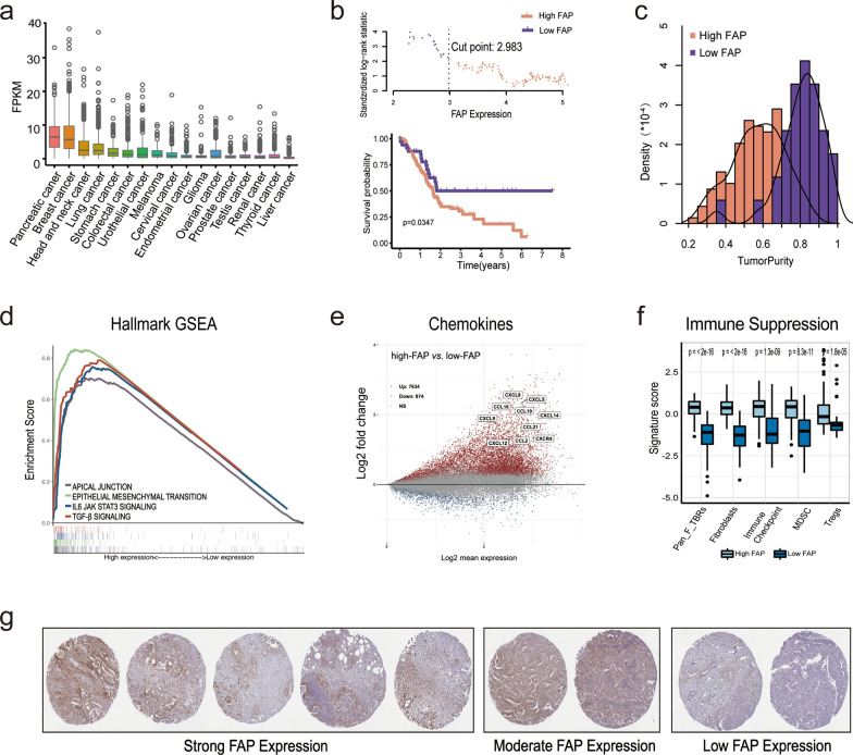
Purpose: The claudin 18.2 (CLDN18.2) antigen is frequently expressed in malignant tumors, including pancreatic ductal adenocarcinoma (PDAC). Although CLDN18.2-targeted CAR-T cells demonstrated some therapeutic efficacy in PDAC patients, further improvement is needed. One of the major obstacles might be the abundant cancer-associated fibroblasts (CAFs) in the PDAC tumor microenvironment (TME). Targeting fibroblast activation protein (FAP), a vital characteristic of CAFs provides a potential way to overcome this obstacle. In this study, we explored the combined antitumor activity of FAP-targeted and CLDN18.2-targeted CAR-T cells against PDAC.

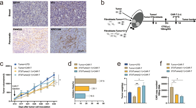
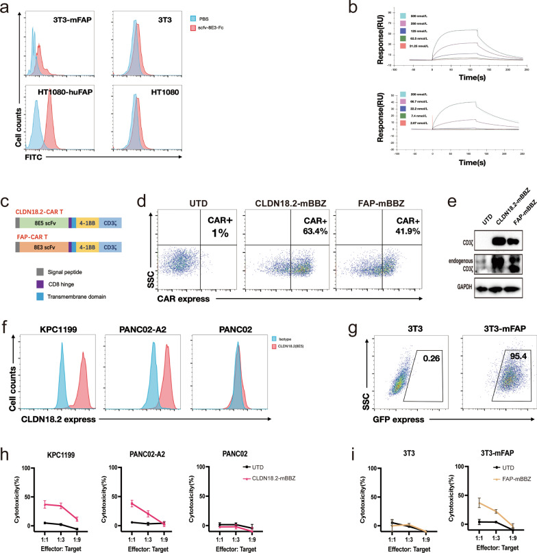
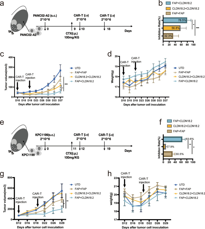
Methods: Novel FAP-targeted CAR-T cells were developed. Sequential treatment of FAP-targeted and CLDN18.2-targeted CAR-T cells as well as the corresponding mechanism were explored in immunocompetent mouse models of PDAC.

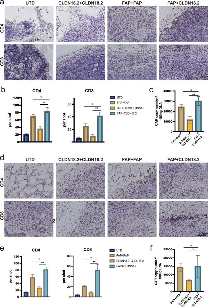
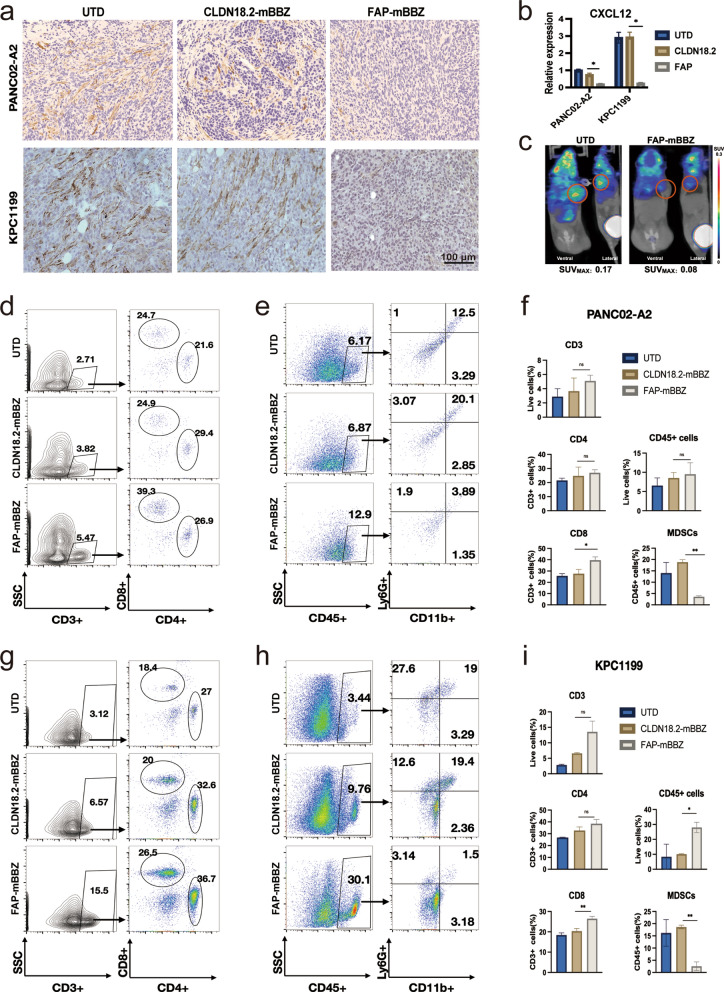
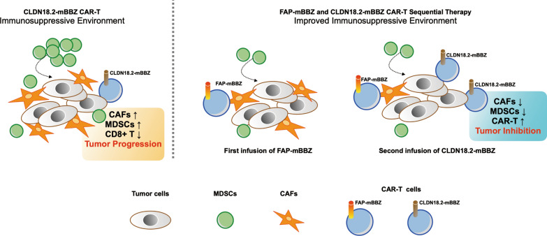
Results: The results indicated that the priorly FAP-targeted CAR-T cells infusion could significantly eliminate CAFs and enhance the anti-PDAC efficacy of subsequently CLDN18.2-targeted CAR-T cells in vivo. Interestingly, we observed that FAP-targeted CAR-T cells could suppress the recruitment of myeloid-derived suppressor cells (MDSCs) and promote the survival of CD8+ T cells and CAR-T cells in tumor tissue.

Conclusion: In summary, our finding demonstrated that FAP-targeted CAR-T cells could increase the antitumor activities of sequential CAR-T therapy via remodeling TME, at least partially through inhibiting MDSCs recruitment. Sequential infusion of FAP-targeted and CLDN18.2-targeted CAR-T cells might be a feasible approach to enhance the clinical outcome of PDAC.


Conflict of interest statement

Dr. Hua Jiang and Dr. Zonghai Li has ownership interests of CAR-T cells relating to this work and is a stockholder of CARsgen Therapeutics, Inc. The other authors declare that they have no conflicts of interest.

Figures

Fig. 1
Fig. 1
Bioinformatics analysis of FAP biological and tumor microenvironment characteristics in pancreatic cancer. a Pan-cancer analysis of FAP expression. b Identification of optimal FAP expression cutoff dividing PDAC cohort. The upper scatter plot shows each cutoff point’s standardized log-rank statistic value. The lower Kaplan-Meier plot shows overall survival for patients divided by the optimal cutoff. cf Biology and TME characteristics in high-FAP vs. low-FAP groups of PDAC cohort. c Density distribution of tumor purity. d Enrichment score of HALLMARK gene-sets related to high FAP expression. e Volcano map of the chemokines. f Immune suppressive characteristics estimated by IOBR. g IHC staining of FAP protein expression in 9 pancreatic cancer samples
Fig. 2
Fig. 2
CAFs impair the antitumor activity of CAR-T cells in vivo. a IHC staining of CAFs in 4T1 and E0771 breast and PANC02 and KPC1199 pancreatic tumor-bearing mice. b-f In vivo experimental of Antigen-Positive E0771 allografts. b Experimental scheme. Antigen-Positive E0771 tumor cells alone or combined with NIH 3T3 fibroblast were in situ inoculated into C57BL/6 mice, treated i.p. injection with CPA, and then given CAR-T cells (i.v.; n = 5 mice per group). c The tumor volume of tumors of each treatment group. d The tumor growth inhibition of each treatment group. e The tumor weight at the end point of the animal experiment. f CAR copy numbers in genomic DNA of residual tumors after therapy were measured by qRT-PCR (TaqMan probe). The images were obtained under original magnification 200×. Scale bars, 100 μm. All data are presented as the mean ± SEM of triplicate experiments. *p < 0.05, **p < 0.01, ***p < 0.001
Fig. 3
Fig. 3
Generation of FAP-targeted CAR-T cells and CLDN18.2-targeted CAR-T cells. a Binding specificity of the anti-FAP antibody to mFAP-transfected 3T3 or huFAP-transfected HT-1080. b Affinity measurements of anti-FAP antibody binding to FAP through BiacoreSurface Plasmon resonance (GE Healthcare). The upper shows its binding to mu-FAP and, the lower shows its binding to hu-FAP. c The construction of 8E3-mBBZ and 8E5-mBBZ CAR-T cells. This construct includes an extracellular antigen recognition region (8E5 targeting CLDN18.2 and 8E3 targeting FAP), a hinge, a transmembrane domain, an intracellular region of mouse 41-BB costimulatory molecules, and a mouse CD3-ζ chain. d The transduction efficiency of FAP-mBBZ and CLDN18.2-BBZ CAR-T CAR on splenic T cells derived from C57BL/6 was determined by flow cytometry. UTD cells served as negative controls. e Western blot of CD3-ζ in CAR-T. Glyceraldehyde 3-phosphate dehydrogenase (GAPDH) served as a loading control. f The expression of CLDN18.2 on KPC1199, PANC02, and PANC02-A2. Cells incubated with a mouse anti-mouse IgG antibody as a negative control. g The establishment of mFAP NIH 3T3 mouse fibroblast cells. NIH 3T3 was transduced by pWPT-GFP-mFAP lentivirus and further sorted by flow cytometry. h-i CAR-T cells were incubated with tumor or fibroblast cells at three effector: target (E: T) ratios for 18 h. Cell lysis was tested using a standard nonradioactive cytotoxicity assay. h Cytotoxic activities of CLDN18.2-mBBZ CAR-T cells on CLDN18.2-positive or CLDN18.2-negative tumor cells. i Cytotoxic activities of FAP-mBBZ CAR-T cells on FAP-positive or FAP-negative fibroblast cells
Fig. 4
Fig. 4
Sequential therapy of FAP-targeted and CLDN18.2-targeted cells dramatically improved antitumor efficacy against CLDN18.2-positive tumor-bearing mice. a-d In vivo experimental design. C57BL/6 mice were injected s.c. with PANC02-A2 cells and allowed to establish for 10 days. Mice were assigned to four experimental groups. The first and second CAR-T cells were injected on day 10 and day 18, respectively (i.v.; n = 5 mice per group). b The tumor growth inhibition of each treatment group. c The volume of tumors of each treatment group. d The body weight of mice of each treatment group. e-h In vivo experimental design. C57BL/6 mice were injected s.c. with KPC1199 cells and allowed to establish for 12 days. Mice were assigned to four experimental groups. The first and second CAR-T cells were injected on day 12 and day 19, respectively (i.v.; n = 5 mice per group). f The tumor growth inhibition of each treatment group. g The volume of tumors of each treatment group. h The body weight of mice of each treatment group. All data are presented as the mean ± SEM of triplicate experiments. *p < 0.05, **p < 0.01, ***p < 0.001
Fig. 5
Fig. 5
Sequential therapy of FAP-targeted and CLDN18.2-targeted CAR-T cells dramatically increase the accumulation of CD8+ T and CAR-T cells. a–c Tumor tissues were harvested from the mice at the end of therapy in the PANC02 model. a The representative images of tumor-infiltrating T cells immunostained with anti-CD4 and anti-CD8 from each treatment group. b Histograms show the quantification of T cells in tumor tissues. c CAR copy numbers in genomic DNA of residual tumors from each treatment group were measured by qRT-PCR (TaqMan probe). df Tumor tissues were harvested from the mice at the end of therapy in the KPC1199 model. d The representative images of tumor-infiltrating T cells immunostained with anti-CD4 and anti-CD8 from each treatment group. e Histograms show the quantification of T cells in tumor tissues. f CAR copy numbers in genomic DNA of residual tumors from each treatment group were measured by qRT-PCR (TaqMan probe). The images were obtained under original magnification 200×. Scale bars, 100 μm. All data are presented as the mean ± SEM of triplicate experiments. *p < 0.05, **p < 0.01
Fig. 6
Fig. 6
FAP-targeted CAR-T cells eliminate CAFs and change tumor microenvironment in vivo. C57BL/6 mice were injected s.c. with PANC02-A2 or KPC1199 cells and treated with CAR-T cells after the tumor was established. To exclude the influence of tumor volume difference by distinct CAR-T cells, tumor tissue was harvested 7 days after treatment. ac Analysis of the CAFs in tumor tissues of PANC02-A2 and KPC1199 allografts. a The representative images of CAFs immunostained with α-SMA in tumor tissues from each treatment group. b q-PCR measured the mRNA expression of CXCL12 in tumor tissues from each treatment group. c PET image labeling FAP by FAPI in PANC02-A2 allografts 7 days after the treatment of FAP-mBBZ or UTD T cells.  d–f  Analysis of the immune cell in tumor tissues of PANC02-A2 allografts. d The representative flow cytometry plots showing the frequencies of tumor-infiltrating CD8+ and CD4+ cells in CD3+ T of each treatment group. e The representative flow cytometry plots showing the frequencies of tumor-infiltrating CD45+ immune cells and MDSCs of each treatment group. f Flow cytometry quantitation of tumor-infiltrating immune cells of each treatment group. gi Analysis of the immune cell in tumor tissues of KPC1199 allografts. g The representative flow cytometry plots showing the frequencies of tumor-infiltrating CD8+ and CD4+ cells in CD3+ T of each treatment group. h The representative flow cytometry plots showing the frequencies of tumor-infiltrating CD45+ immune cells and MDSCs of each treatment group. i Flow cytometry quantitation of tumor-infiltrating immune cells of each treatment group. The images were obtained under original magnification 200×. Scale bars, 100 μm. All data are presented as the mean ± SEM of triplicate experiments. *p < 0.05, **p < 0.01, *** p < 0.001
Fig. 7
Fig. 7
Schema: Sequential Therapy enhances the antitumor ability of CAR-T by suppressing the infiltration of MDSCs. Left: CAFs are crucial in TME, forming an immune surveillance barrier and perpetuating tumor-promoting. The CAFs could also promote recruit monocytes from the bone marrow, such as MDSCs, to form a tumor-suppressing microenvironment, suppressing CAR-T cell function. Right: FAP-targeted CAR-T cells eliminate CAFs via specific recognition of FAP on CAFs in the tumor microenvironment. Further, CLDN18.2-targeted CAR-T cells could also increase cytotoxic T cells and inhibit the recruiting of MDSCs. With an improved immune suppressive microenvironment, the antitumor effect of the sequential infusion of CLDN18.2-targeted CAR-T cells were enhanced

Similar articles

Cited by

References

    1. Mizrahi JD, Surana R, Valle JW, Shroff RT. Pancreatic cancer. Lancet. 2020;395(10242):2008–20. doi: 10.1016/S0140-6736(20)30974-0. - DOI - PubMed
    1. Sung H, Ferlay J, Siegel RL, Laversanne M, Soerjomataram I, Jemal A, et al. Global Cancer Statistics 2020: GLOBOCAN estimates of incidence and mortality worldwide for 36 cancers in 185 countries. CA Cancer J Clin. 2021;71(3):209–49. doi: 10.3322/caac.21660. - DOI - PubMed
    1. Neelapu SS, Locke FL, Bartlett NL, Lekakis LJ, Miklos DB, Jacobson CA, et al. Axicabtagene ciloleucel CAR T-Cell therapy in refractory large B-Cell lymphoma. N Engl J Med. 2017;377(26):2531–44. doi: 10.1056/NEJMoa1707447. - DOI - PMC - PubMed
    1. Jiang H, Shi Z, Wang P, Wang C, Yang L, Du G, et al. Claudin18.2-Specific chimeric Antigen receptor Engineered T cells for the treatment of gastric Cancer. J Natl Cancer Inst. 2019;111(4):409–18. doi: 10.1093/jnci/djy134. - DOI - PubMed
    1. Qi C, Gong J, Li J, Liu D, Qin Y, Ge S, et al. Claudin18.2-specific CAR T cells in gastrointestinal cancers: phase 1 trial interim results. Nat Med. 2022;28(6):1189–98. doi: 10.1038/s41591-022-01800-8. - DOI - PMC - PubMed

Publication types

Substances