

Longitudinal Behavior of Left-Ventricular Strain in Fetal Growth Restriction
- PMID: 37046470
- PMCID: PMC10093576
- DOI: 10.3390/diagnostics13071252
Longitudinal Behavior of Left-Ventricular Strain in Fetal Growth Restriction
Abstract
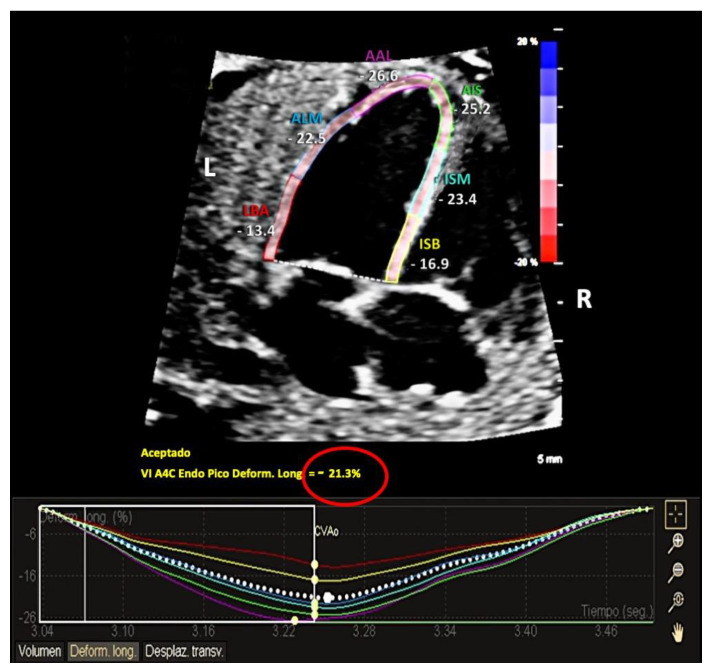
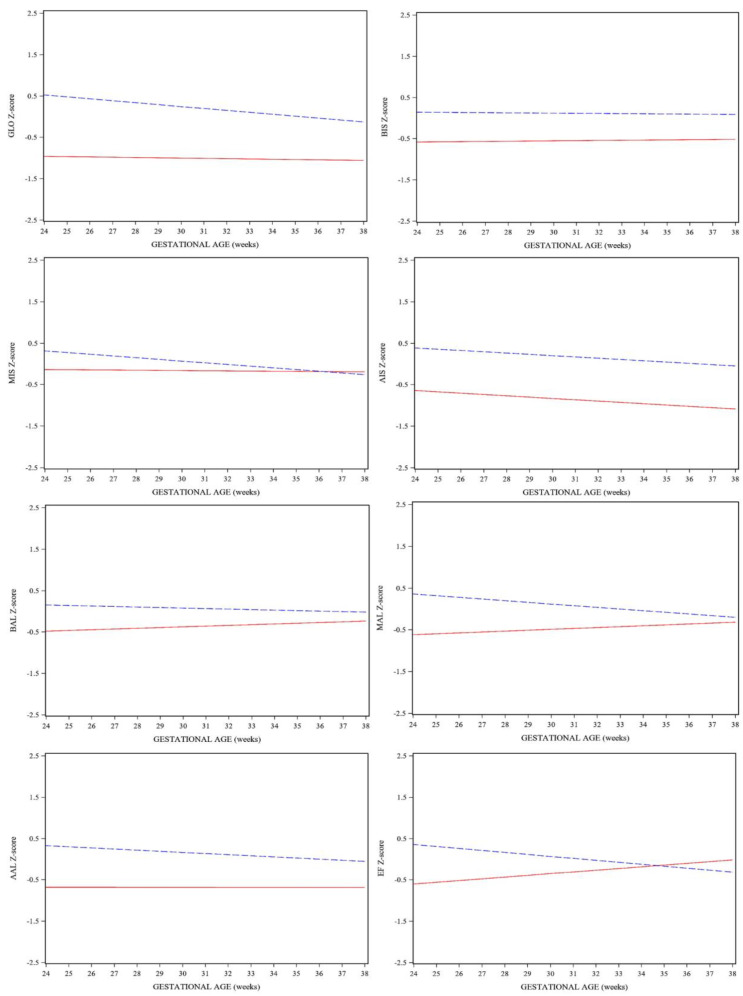
Fetal growth restriction (FGR) is associated with an increased risk of adverse outcomes resulting from adaptive cardiovascular changes in conditions of placental insufficiency, leading to cardiac deformation and dysfunction, which can be evaluated with 2D speckle tracking echocardiography (2D-STE). The aim of the present study was to evaluate whether reduced fetal growth is associated with cardiac left-ventricle (LV) dysfunction, using 2D-STE software widely used in postnatal echocardiography. A prospective longitudinal cohort study was performed, and global (GLO) and segmental LV longitudinal strain was measured offline and compared between FGR and appropriate-for-gestational-age (AGA) fetuses throughout gestation. All cases of FGR fetuses were paired 1:2 to AGA fetuses, and linear mixed model analysis was performed to compare behavior differences between groups throughout pregnancy. Our study shows LV fetal longitudinal strain in FGR and AGA fetuses differed upon diagnosis and behaved differently throughout gestation. FGR fetuses had lower LV strain values, both global and segmental, in comparison to AGA, suggesting subclinical cardiac dysfunction. Our study provides more data regarding fetal cardiac function in cases of placental dysfunction, as well as highlights the potential use of 2D-STE in the follow-up of cardiac function in these fetuses.
Keywords: 2D speckle tracking; aCMQ-QLab; fetal echocardiography; fetal growth restriction; small for gestational age; strain.
Conflict of interest statement
Elisa Llurba declares lecture fees from Cook, Viñas, and ROCHE diagnostics.
Figures
Similar articles
-
Fetal Left Ventricle Function Evaluated by Two-Dimensional Speckle-Tracking Echocardiography across Clinical Stages of Severity in Growth-Restricted Fetuses.Diagnostics (Basel). 2024 Mar 5;14(5):548. doi: 10.3390/diagnostics14050548. Diagnostics (Basel). 2024. PMID: 38473020 Free PMC article.
-
Value of two-dimensional speckle-tracking echocardiography in evaluation of cardiac function in small fetuses.Quant Imaging Med Surg. 2024 Dec 5;14(12):8155-8166. doi: 10.21037/qims-24-794. Epub 2024 Oct 24. Quant Imaging Med Surg. 2024. PMID: 39698698 Free PMC article.
-
Two-dimensional Speckle tracking echocardiography in Fetal Growth Restriction: a systematic review.Eur J Obstet Gynecol Reprod Biol. 2020 Nov;254:87-94. doi: 10.1016/j.ejogrb.2020.08.052. Epub 2020 Sep 2. Eur J Obstet Gynecol Reprod Biol. 2020. PMID: 32950891
-
Myocardial deformation analysis in late-onset small-for-gestational-age and growth-restricted fetuses using two-dimensional speckle tracking echocardiography: a prospective cohort study.J Perinat Med. 2021 Sep 17;50(3):305-312. doi: 10.1515/jpm-2021-0162. Print 2022 Mar 28. J Perinat Med. 2021. PMID: 34529908
-
Gestational Age-Adjusted Reference Ranges for Fetal Left Ventricle Longitudinal Strain by Automated Cardiac Motion Quantification between 24 and 37 Weeks' Gestation.Fetal Diagn Ther. 2022;49(7-8):311-320. doi: 10.1159/000527120. Epub 2022 Sep 20. Fetal Diagn Ther. 2022. PMID: 36126644
Cited by
-
Fetal Left Ventricle Function Evaluated by Two-Dimensional Speckle-Tracking Echocardiography across Clinical Stages of Severity in Growth-Restricted Fetuses.Diagnostics (Basel). 2024 Mar 5;14(5):548. doi: 10.3390/diagnostics14050548. Diagnostics (Basel). 2024. PMID: 38473020 Free PMC article.
-
Evaluating the Impact of Maternal Lipid Profiles on Fetal Cardiac Function at Mid-Gestation: An Observational Study.Clin Pract. 2024 Nov 27;14(6):2590-2600. doi: 10.3390/clinpract14060204. Clin Pract. 2024. PMID: 39727792 Free PMC article.
-
Fetal heart quantification ultrasound technology for the quantitative analysis of fetal cardiac morphology and function in hypertensive disorders of pregnancy.Quant Imaging Med Surg. 2025 Apr 1;15(4):3517-3531. doi: 10.21037/qims-24-1553. Epub 2025 Mar 28. Quant Imaging Med Surg. 2025. PMID: 40235737 Free PMC article.
-
Value of two-dimensional speckle-tracking echocardiography in evaluation of cardiac function in small fetuses.Quant Imaging Med Surg. 2024 Dec 5;14(12):8155-8166. doi: 10.21037/qims-24-794. Epub 2024 Oct 24. Quant Imaging Med Surg. 2024. PMID: 39698698 Free PMC article.
References
-
- Hanchate N., Ramani S., Mathpati C.S., Dalvi V.H. Biomass Gasification Using Dual Fluidized Bed Gasification Systems: A Review. J. Clean. Prod. 2021;280:123148. doi: 10.1016/j.jclepro.2020.123148. - DOI
-
- Crispi F., Hernandez-Andrade E., Pelsers M.M.A.L., Plasencia W., Benavides-Serralde J.A., Eixarch E., Le Noble F., Ahmed A., Glatz J.F.C., Nicolaides K.H., et al. Cardiac Dysfunction and Cell Damage across Clinical Stages of Severity in Growth-Restricted Fetuses. Am. J. Obstet. Gynecol. 2008;199:254.e1–254.e8. doi: 10.1016/j.ajog.2008.06.056. - DOI - PubMed
LinkOut - more resources
Full Text Sources
Research Materials
Miscellaneous
