

Spinal Schwannomatosis Mimicking Metastatic Extramedullary Spinal Tumor
- PMID: 37046472
- PMCID: PMC10092950
- DOI: 10.3390/diagnostics13071254
Spinal Schwannomatosis Mimicking Metastatic Extramedullary Spinal Tumor
Abstract
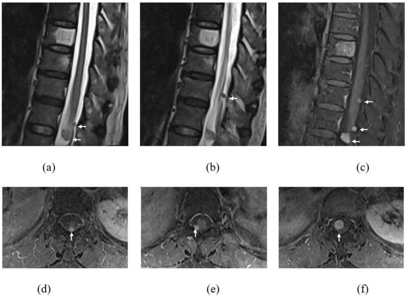
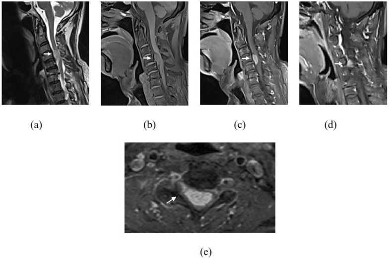
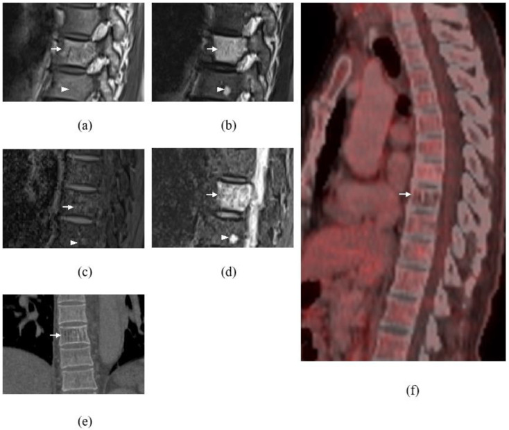
Intradural extramedullary (IDEM) tumors are the most commonly observed intraspinal tumors, comprising over 60% of tumors found within the spinal canal, and the vast majority of these lesions are benign lesions. IDEM metastases are rare, but if they occur, they commonly manifest as leptomeningeal disease, secondary to drop lesions from intracranial metastases from adenocarcinomas of the lung, prostate cancer, breast cancer, melanoma, or rarely, as a result of lymphomas. The purely non-neurogenic origin of IDEM metastases is rare. Herein, we describe a patient with a previous history of treated colon cancer who presented with a progressive neurological deficit and whose imaging revealed multiple intradural, extramedullary and osseous lesions at the cervical and thoracolumbar spines. With the previous known primary and multiplicity of the lesions, an initial diagnosis of spinal metastasis was made, But it was proven to be schwannoma on histology. We emphasize the diagnostic dilemma in this case and the importance of detecting subtle imaging findings, which may be helpful to differentiate between metastatic disease and a second primary tumor.
Keywords: extramedullary; hemangioma; intradural; metastases; schwannoma.
Conflict of interest statement
The authors declare no conflict of interest.
Figures
Similar articles
-
Myelopathy from Intradural Extramedullary Metastasis as an Initial Presentation of Metastatic Melanoma.Cureus. 2018 May 22;10(5):e2668. doi: 10.7759/cureus.2668. Cureus. 2018. PMID: 30042919 Free PMC article.
-
Intradural Extramedullary Metastatic Conjunctival Malignant Melanoma.World Neurosurg. 2020 Jun;138:444-448. doi: 10.1016/j.wneu.2020.03.078. Epub 2020 Mar 23. World Neurosurg. 2020. PMID: 32217182
-
Spinal metastasis of parotid acinic cell carcinoma followed by intradural extramedullary recurrence: illustrative case.J Neurosurg Case Lessons. 2021 Dec 20;2(25):CASE21591. doi: 10.3171/CASE21591. eCollection 2021 Dec 20. J Neurosurg Case Lessons. 2021. PMID: 35855290 Free PMC article.
-
Intradural extramedullary cavernoma of a lumbar nerve root mimicking neurofibroma. A report of a rare case and the differential diagnosis.Spine J. 2014 Dec 1;14(12):e1-7. doi: 10.1016/j.spinee.2014.08.447. Epub 2014 Sep 6. Spine J. 2014. PMID: 25200326 Review.
-
Primary Intradural Extramedullary Sporadic Spinal Hemangioblastomas: Case Report and Systematic Review.World Neurosurg. 2021 Aug;152:84-94. doi: 10.1016/j.wneu.2021.05.105. Epub 2021 Jun 1. World Neurosurg. 2021. PMID: 34087464
Cited by
-
Animal models of brain and spinal cord metastases of NSCLC established using a brain stereotactic instrument.Heliyon. 2024 Jan 20;10(3):e24809. doi: 10.1016/j.heliyon.2024.e24809. eCollection 2024 Feb 15. Heliyon. 2024. PMID: 38318004 Free PMC article.
-
Solitary intradural extramedullary recurrence of testicular diffuse large B-cell lymphoma mimicking a schwannoma: illustrative case.J Neurosurg Case Lessons. 2025 Apr 21;9(16):CASE24830. doi: 10.3171/CASE24830. Print 2025 Apr 21. J Neurosurg Case Lessons. 2025. PMID: 40258327 Free PMC article.
References
-
- Tieng F.Y.F., Baharudin R., Abu N., Mohd Yunos R.I., Lee L.H., Ab Mutalib N.S. Single Cell Transcriptome in Colorectal Cancer-Current Updates on Its Application in Metastasis, Chemoresistance and the Roles of Circulating Tumor Cells. Front. Pharmacol. 2020;11:135. doi: 10.3389/fphar.2020.00135. PMCID: PMC7056698. - DOI - PMC - PubMed
-
- Thanuja M., Azyani Y. Osteofibrous Dysplasia-Like Adamantinoma Mimicking Bone Metastasis in A Patient with Rectosigmoid Cancer. Malays. J. Med. Health Sci. 2021;17:200–202.
LinkOut - more resources
Full Text Sources
