Simultaneous Super-Resolution and Classification of Lung Disease Scans
- PMID: 37046537
- PMCID: PMC10093568
- DOI: 10.3390/diagnostics13071319
Simultaneous Super-Resolution and Classification of Lung Disease Scans
Abstract
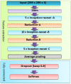
Acute lower respiratory infection is a leading cause of death in developing countries. Hence, progress has been made for early detection and treatment. There is still a need for improved diagnostic and therapeutic strategies, particularly in resource-limited settings. Chest X-ray and computed tomography (CT) have the potential to serve as effective screening tools for lower respiratory infections, but the use of artificial intelligence (AI) in these areas is limited. To address this gap, we present a computer-aided diagnostic system for chest X-ray and CT images of several common pulmonary diseases, including COVID-19, viral pneumonia, bacterial pneumonia, tuberculosis, lung opacity, and various types of carcinoma. The proposed system depends on super-resolution (SR) techniques to enhance image details. Deep learning (DL) techniques are used for both SR reconstruction and classification, with the InceptionResNetv2 model used as a feature extractor in conjunction with a multi-class support vector machine (MCSVM) classifier. In this paper, we compare the proposed model performance to those of other classification models, such as Resnet101 and Inceptionv3, and evaluate the effectiveness of using both softmax and MCSVM classifiers. The proposed system was tested on three publicly available datasets of CT and X-ray images and it achieved a classification accuracy of 98.028% using a combination of SR and InceptionResNetv2. Overall, our system has the potential to serve as a valuable screening tool for lower respiratory disorders and assist clinicians in interpreting chest X-ray and CT images. In resource-limited settings, it can also provide a valuable diagnostic support.
Keywords: Coronavirus; chest X-ray radiographs; convolutional neural network; image super-resolution; multi-class SVM.
Conflict of interest statement
The authors declare no conflict of interest.
Figures


References
-
- Cruz A.A. Global Surveillance, Prevention and Control of Chronic Respiratory Diseases: A Comprehensive Approach. World Health Organization; Geneva, Switzerland: 2007.
-
- WHO . World Health Organization Coronavirus Disease 2019 (COVID-19) Situation Report. World Health Organization; Geneva, Switzerland: 2020.
-
- Rahaman M.M., Li C., Yao Y., Kulwa F., Rahman M.A., Wang Q., Qi S., Kong F., Zhu X., Zhao X. Identification of COVID-19 samples from chest X-Ray images using deep learning: A comparison of transfer learning approaches. J. X-ray Sci. Technol. 2020;28:821–839. doi: 10.3233/XST-200715. - DOI - PMC - PubMed
LinkOut - more resources
Full Text Sources
Research Materials
Miscellaneous

