Deep Learning-Based Algorithm for Automatic Detection of Pulmonary Embolism in Chest CT Angiograms
- PMID: 37046542
- PMCID: PMC10093638
- DOI: 10.3390/diagnostics13071324
Deep Learning-Based Algorithm for Automatic Detection of Pulmonary Embolism in Chest CT Angiograms
Abstract
Purpose: Since the prompt recognition of acute pulmonary embolism (PE) and the immediate initiation of treatment can significantly reduce the risk of death, we developed a deep learning (DL)-based application aimed to automatically detect PEs on chest computed tomography angiograms (CTAs) and alert radiologists for an urgent interpretation. Convolutional neural networks (CNNs) were used to design the application. The associated algorithm used a hybrid 3D/2D UNet topology. The training phase was performed on datasets adequately distributed in terms of vendors, patient age, slice thickness, and kVp. The objective of this study was to validate the performance of the algorithm in detecting suspected PEs on CTAs.
Methods: The validation dataset included 387 anonymized real-world chest CTAs from multiple clinical sites (228 U.S. cities). The data were acquired on 41 different scanner models from five different scanner makers. The ground truth (presence or absence of PE on CTA images) was established by three independent U.S. board-certified radiologists.
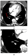
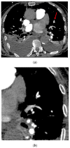
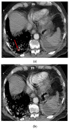
Results: The algorithm correctly identified 170 of 186 exams positive for PE (sensitivity 91.4% [95% CI: 86.4-95.0%]) and 184 of 201 exams negative for PE (specificity 91.5% [95% CI: 86.8-95.0%]), leading to an accuracy of 91.5%. False negative cases were either chronic PEs or PEs at the limit of subsegmental arteries and close to partial volume effect artifacts. Most of the false positive findings were due to contrast agent-related fluid artifacts, pulmonary veins, and lymph nodes.
Conclusions: The DL-based algorithm has a high degree of diagnostic accuracy with balanced sensitivity and specificity for the detection of PE on CTAs.
Keywords: artificial intelligence; chest CT; computed tomography angiography; deep learning tool; pulmonary embolism.
Conflict of interest statement
P.A.G. and B.D.M. have no conflict of interest. D.S.C. and P.D.C. own stock options in Avicenna.AI. A.A., S.Q., M.T., M.M. and Y.C. are employees of Avicenna.AI.
Figures


References
-
- Donato A.A., Scheirer J.J., Atwell M.S., Gramp J., Duszak R., Jr. Clinical outcomes in patients with suspected acute pulmonary embolism and negative helical computed tomographic results in whom anticoagulation was withheld. Arch. Intern. Med. 2003;163:2033–2038. doi: 10.1001/archinte.163.17.2033. - DOI - PubMed

