Extravascular Ultrasound (EVUS) to Assess the Results of Peripheral Endovascular Procedures
- PMID: 37046574
- PMCID: PMC10093749
- DOI: 10.3390/diagnostics13071356
Extravascular Ultrasound (EVUS) to Assess the Results of Peripheral Endovascular Procedures
Abstract
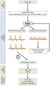
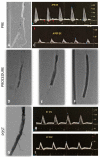
Contrast arteriography (CA) is considered the gold standard to evaluate any phase in peripheral arterial disease (PAD) interventions, from diagnostics to final results. Nevertheless, duplex ultrasonography (DUS) mostly used for the pre/postoperative phase and follow-up control, could be a potential intraoperative adjunctive imaging tool to assess the effects of endovascular revascularization in patients with iliac and femoropopliteal lesions. The PAD "duplex-assisted" protocol includes a preoperative DUS control followed by an intraoperative and a postoperative control. The most important parameters are pulsed doppler spectral analysis and waveform changes, which are impossible to detect with intravascular ultrasound (IVUS). By using a similar acronym, the intraoperative DUS has been previously described as extravascular ultrasound (EVUS). B-mode imaging, color flow, and peak systolic velocity (PSV) are considered. EVUS could be very useful to evaluate the effects of endovascular treatment, mainly in cases of unclear CAs, severe calcifications and/or dissections. In the context of the "leaving nothing behind" strategy, EVUS can drive the physician to evaluate the absence of flow-limiting dissections and decide which target lesion should be treated with antirestenotic therapy, further vessel preparation, or stenting. The EVUS protocol could be a safe and feasible option to improve the completion assessment of endovascular PAD treatment. A better ultrasound waveform is a sign of improved luminal gain and compliance, which is extremely important to finalize the results of new peripheral device technology, such as intravascular lithotripsy.
Keywords: EVUS; PAD; angiography; duplex ultrasound; lithotripsy.
Conflict of interest statement
Stefano Fazzini has a consulting agreement with Shockwave Medical.
Figures


References
-
- Polak J.F., Karmel M.I., Mannick J.A., O’Leary D.H., Donaldson M.C., Whittemore A.D. Determination of the extent of lower-extremity peripheral arterial disease with color-assisted duplex sonography: Comparison with angiography. Am. J. Roentgenol. 1990;155:1085–1089. doi: 10.2214/ajr.155.5.2120939. - DOI - PubMed
-
- Karacagil S., Löfberg A., Granbo A., Lörelius L., Bergqvist D. Value of Duplex scanning in evaluation of crural and foot arteries in limbs with severe lower limb ischaemia—A prospective comparison with angiography. Eur. J. Vasc. Endovasc. Surg. 1996;12:300–303. doi: 10.1016/S1078-5884(96)80248-6. - DOI - PubMed
-
- Dougherty M.J., Hallett J.W., Jr., Naessens J.M., Bower T.C., Cherry K.J., Gloviczki P., James E.M. Optimizing technical success of renal revascularization: The impact of intraoperative color-flow duplex ultrasonography. J. Vasc. Surg. 1993;17:849–857. doi: 10.1016/0741-5214(93)90034-J. - DOI - PubMed
LinkOut - more resources
Full Text Sources

