. 2023 Apr 1;15(7):2118.
doi: 10.3390/cancers15072118.

α-Mangostin Promotes In Vitro and In Vivo Degradation of Androgen Receptor and AR-V7 Splice Variant in Prostate Cancer Cells

Affiliations

α-Mangostin Promotes In Vitro and In Vivo Degradation of Androgen Receptor and AR-V7 Splice Variant in Prostate Cancer Cells

Mirielle C Nauman et al. Cancers (Basel). .

Abstract

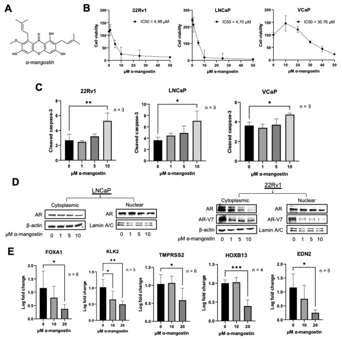
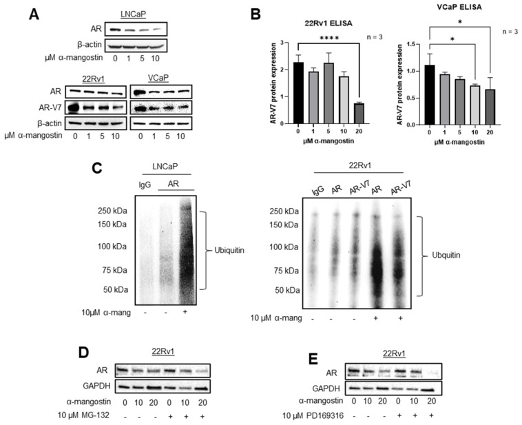
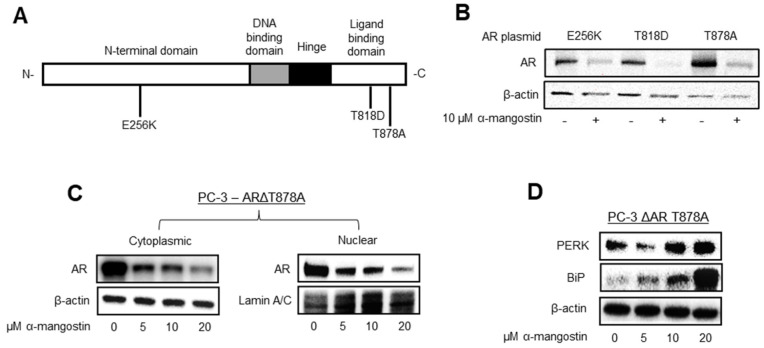
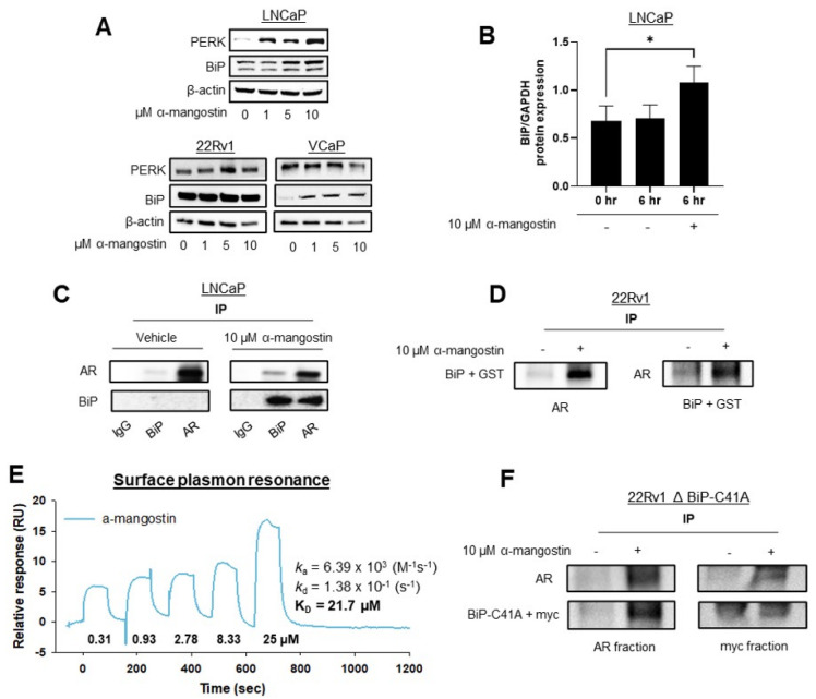
A major limitation of current prostate cancer pharmacotherapy approaches is the inability of these compounds to target androgen receptor variants or mutants that develop during prostate cancer progression. The demand for novel therapeutics to prevent, slow, and treat prostate cancer is significant because FDA approved anti-androgens are associated with adverse events and can eventually drive drug-resistant prostate cancer. This study evaluated α-mangostin for its novel ability to degrade the androgen receptor and androgen receptor variants. α-Mangostin is one of more than 70 isoprenylated xanthones isolated from Garcinia mangostana that we have been evaluating for their anticancer potential. Prostate cancer cells treated with α-mangostin exhibited decreased levels of wild-type and mutated androgen receptors. Immunoblot, immunoprecipitation, and transfection experiments demonstrated that the androgen receptor was ubiquitinated and subsequently degraded via the proteasome, which we hypothesize occurs with the assistance of BiP, an ER chaperone protein that we have shown to associate with the androgen receptor. We also evaluated α-mangostin for its antitumor activity and promotion of androgen receptor degradation in vivo. In summary, our study demonstrates that androgen receptor degradation occurs through the novel activation of BiP and suggests a new therapeutic approach for prostate cancer.

Keywords: AR-V7; BiP; GRP78; Garcinia mangostana; androgen receptor; mangosteen; prostate cancer; xanthone; α-mangostin.


Conflict of interest statement

The authors declare no conflict of interest.

Figures

Figure 1
Figure 1
α-Mangostin promotes apoptosis and inhibits nuclear translocation. (A) Structure of α-mangostin. (B) Cells were treated with α-mangostin and cell viability was determined by MTT assays. (C) Cells were treated with 0–20 μM α-mangostin for 24 h and cleaved caspase-3 levels were analyzed by ELISA. 30 µg of protein from cell lysates were used. (D) Nuclear and cytoplasmic fractions from cells treated with α-mangostin for 24 h were analyzed for androgen receptor expression by Western blot. (E) AR- and AR-V7-regulated genes were analyzed by qPCR in 22Rν1 cells treated with α-mangostin for 24 h. * p ≤ 0.05, ** p ≤ 0.01, and *** p ≤ 0.001. The uncropped blots are shown in File S1.
Figure 2
Figure 2
α-Mangostin promotes the ubiquitination and degradation of AR and AR-V7 via the proteasome. (A) AR and AR-V7 expression was evaluated in α-mangostin-treated PCa cells, including LNCaP, 22Rν1, and VCaP cells. (B) 22Rν1 and VCaP cells were treated with α-mangostin for 24 h and then whole cell lysates were evaluated for their expression of total AR-V7 through ELISA assays. (C) AR proteins from LNCaP and AR and AR-V7 proteins from 22Rν1 were immunoprecipitated with either anti-AR or anti-AR-V7 antibodies. Ubiquitinated AR and AR-V7 were analyzed by Western blot. (D) AR expression was analyzed after either individual or co-treatments of α-mangostin and/or MG-132. (E) AR expression was analyzed after either individual or co-treatments of α-mangostin and/or PD169316. * p ≤ 0.05 and **** p ≤ 0.0001. The uncropped blots are shown in File S1.
Figure 3
Figure 3
α-Mangostin decreases mutant AR expression and promotes the expression of BiP. (A) Structure of AR with single amino acid point mutations. (B) AR expression was evaluated in α-mangostin-treated PC-3 cells that had been transfected with 0.5–1 µg of ΔAR E256K, T818D, or T878A plasmid. (C) AR expression was evaluated in PC-3 cells treated with varying concentrations of α-mangostin that had been transfected with 0.5 µg of ΔAR T878A plasmid. (C) AR expression was evaluated in the nuclear and cytoplasmic fractions of cell lysates from α-mangostin-treated PC-3 cells transfected with 0.5 µg of ΔAR T878A plasmid. (D) Westerns of PERK and BiP expression in PC-3 cells transfected with ΔAR T878A cDNA after 24-h treatments. The uncropped blots are shown in File S1.
Figure 4
Figure 4
α-Mangostin increased BiP expression, promoted a protein-protein interaction between AR and BiP, and binds to BiP. (A) Westerns of PERK and BiP expression after 24-h treatments. (B) Quantification of Western blots evaluating BiP expression after 6-h treatments with α-mangostin in LNCaP cells. (C) Immunoprecipitation performed in LNCaP cells after 24-h treatments of 10 µM α-mangostin. The AR-protein and BiP-protein that were pulled down were both analyzed for the presence of AR and BiP. (D) 22Rν1 cells were transfected with 15 μg of BiP + GST cDNA. After 24 h, cells were treated with 10 μM α-mangostin and both AR-protein and BiP + GST protein were immunoprecipitated and analyzed for the presence of AR or GST. (E) SPR analysis of the His-GST-BiP protein revealed that α-mangostin bound to BiP. (F) BiP plasmid containing a C41A mutation (15 μg) was transfected into 22Rν1 cells. Immunoprecipitation analyses were repeated, and lysates were analyzed for the presence of AR or myc. * p ≤ 0.05. The uncropped blots are shown in File S1.
Figure 5
Figure 5
α-Mangostin is more effective Than bicalutamide at reducing tumor size and slowing cancer growth in a 22Rν1 xenograft study. (A) The body weights of three cohorts (control, bicalutamide, or α-mangostin) were measured weekly for the duration of the study. (B) Tumor volume measurements were taken five times during the study using a digital caliper. (C) After the animal sacrifice, mouse tumors were photographed. (D) Mouse plasma was collected 1 h after dosing on the day of sacrifice. 50 μL of either the PSA standard or plasma sample was added to a pre-treated ELISA plate, and absorbance readings were taken. * p ≤ 0.05.
Figure 6
Figure 6
α-Mangostin decreased AR and AR-V7 protein expression and related genes in vivo. (A) 20 mg tumor tissue was homogenized in T-PER buffer and protein was extracted. 15 μg protein was loaded, and AR and AR-V7 expressions were detected via Western blot. (B) Immunohistochemistry was performed on 30 μg tumor tissue samples, and an AR antibody was used to stain the slides. (C) The expression of AR and AR-V7-regulated genes was evaluated in RNA extracted from tumor tissues through qPCR. * p ≤ 0.05. The uncropped blots are shown in File S1.

Similar articles

Cited by

References

    1. Rawla P. Epidemiology of Prostate Cancer. World J. Oncol. 2019;10:63–89. doi: 10.14740/wjon1191. - DOI - PMC - PubMed
    1. Bray F., Ferlay J., Soerjomataram I., Siegel R.L., Torre L.A., Jemal A. Global cancer statistics 2018: GLOBOCAN estimates of incidence and mortality worldwide for 36 cancers in 185 countries. CA Cancer J. Clin. 2018;68:394–424. doi: 10.3322/caac.21492. - DOI - PubMed
    1. Sandhu G.S., Nepple K.G., Tanagho Y.S., Andriole G.L. Prostate cancer chemoprevention. Semin. Oncol. 2013;40:276–285. doi: 10.1053/j.seminoncol.2013.04.003. - DOI - PubMed
    1. Chhabra G., Singh C.K., Ndiaye M.A., Fedorowicz S., Molot A., Ahmad N. Prostate cancer chemoprevention by natural agents: Clinical evidence and potential implications. Cancer Lett. 2018;422:9–18. doi: 10.1016/j.canlet.2018.02.025. - DOI - PMC - PubMed
    1. Jin H.J., Kim J., Yu J. Androgen receptor genomic regulation. Transl. Androl. Urol. 2013;2:157–177. doi: 10.3978/j.issn.2223-4683.2013.09.01. - DOI - PMC - PubMed