. 2023 Apr 5;15(7):2150.
doi: 10.3390/cancers15072150.

Development and Evaluation of MR-Based Radiogenomic Models to Differentiate Atypical Lipomatous Tumors from Lipomas

Affiliations

Development and Evaluation of MR-Based Radiogenomic Models to Differentiate Atypical Lipomatous Tumors from Lipomas

Sarah C Foreman et al. Cancers (Basel). .

Abstract

Background: The aim of this study was to develop and validate radiogenomic models to predict the MDM2 gene amplification status and differentiate between ALTs and lipomas on preoperative MR images.

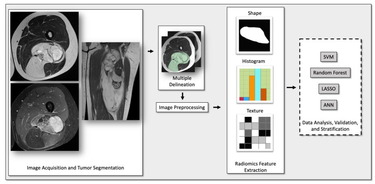
Methods: MR images were obtained in 257 patients diagnosed with ALTs (n = 65) or lipomas (n = 192) using histology and the MDM2 gene analysis as a reference standard. The protocols included T2-, T1-, and fat-suppressed contrast-enhanced T1-weighted sequences. Additionally, 50 patients were obtained from a different hospital for external testing. Radiomic features were selected using mRMR. Using repeated nested cross-validation, the machine-learning models were trained on radiomic features and demographic information. For comparison, the external test set was evaluated by three radiology residents and one attending radiologist.

Results: A LASSO classifier trained on radiomic features from all sequences performed best, with an AUC of 0.88, 70% sensitivity, 81% specificity, and 76% accuracy. In comparison, the radiology residents achieved 60-70% accuracy, 55-80% sensitivity, and 63-77% specificity, while the attending radiologist achieved 90% accuracy, 96% sensitivity, and 87% specificity.

Conclusion: A radiogenomic model combining features from multiple MR sequences showed the best performance in predicting the MDM2 gene amplification status. The model showed a higher accuracy compared to the radiology residents, though lower compared to the attending radiologist.

Keywords: MRI; machine learning; radiology; radiomics; soft-tissue sarcomas.


Conflict of interest statement

The authors declare no conflict of interest.

Figures

Figure 1
Figure 1
Subject selection flowchart. ALT = atypical lipomatous tumor; MDM2 = murine double minute.
Figure 2
Figure 2
Radiomic workflow. Abbreviations: SVM, support vector machine; LASSO, least absolute shrinkage and selection operator; ANN, artificial neural network.
Figure 3
Figure 3
Lipomatous tumor in the medial right thigh, encasing the gracilis muscle (G). (A) The axial T2-weighted and (B) axial T1-weighted MR images show a large heterogeneous tumor with thick septa. (C) Septal contrast enhancement on the coronal T1-weighted images with fat saturation. (D) The machine-learning algorithm classified the tumor as an ALT with a probability of 99.8%. This diagnosis was confirmed by pathology and immunohistochemistry after surgical resection.
Figure 4
Figure 4
Axial T2-weighted (A) and T1-weighted (B) MR images showing a well-defined intramuscular lipomatous tumor (lipoma) in the right posterior thigh without significant contrast enhancement on the axial T1-weighted image with fat saturation (C). (D) The machine-learning model classified this tumor as a lipoma (probability of 97.8%). This was in accordance with the diagnosis made by the radiology residents and the attending radiologist.
Figure 5
Figure 5
Sagittal T2-weighted (A) and axial T1-weighted (B) MR images of a lipomatous tumor located subcutaneously, anteromedial to the right proximal tibia. (C) A sagittal T1-weighted image with fat saturation shows a moderate septal contrast enhancement. All radiology residents classified this tumor as a lipoma, while the attending radiologist classified this tumor as an ALT. (D) The machine-learning algorithm also classified this tumor as an ALT with a probability of 71.6%. The diagnosis of an ALT was confirmed by pathology after surgical resection.

References

    1. Johnson C.N., Ha A.S., Chen E., Davidson D. Lipomatous Soft-tissue Tumors. J. Am. Acad. Orthop. Surg. 2018;26:779–788. doi: 10.5435/JAAOS-D-17-00045. - DOI - PubMed
    1. Dalal K.M., Antonescu C.R., Singer S. Diagnosis and management of lipomatous tumors. J. Surg. Oncol. 2008;97:298–313. doi: 10.1002/jso.20975. - DOI - PubMed
    1. Myhre-Jensen O., Kaae S., Madsen E.H., Sneppen O. Histopathological grading in soft-tissue tumours. Relation to survival in 261 surgically treated patients. Acta Pathol. Microbiol. Immunol. Scand. A. 1983;91:145–150. - PubMed
    1. Rydholm A., Berg N.O. Size, site and clinical incidence of lipoma. Factors in the differential diagnosis of lipoma and sarcoma. Acta Orthop. Scand. 1983;54:929–934. doi: 10.3109/17453678308992936. - DOI - PubMed
    1. Fletcher C.D. The evolving classification of soft tissue tumours: An update based on the new WHO classification. Histopathology. 2006;48:3–12. doi: 10.1111/j.1365-2559.2005.02284.x. - DOI - PubMed