

New Automated Method for Lung Functional Volumes Delineation with Lung Perfusion PET/CT Imaging
- PMID: 37046827
- PMCID: PMC10093378
- DOI: 10.3390/cancers15072166
New Automated Method for Lung Functional Volumes Delineation with Lung Perfusion PET/CT Imaging
Abstract
Background: Gallium-68 lung perfusion PET/CT is an emerging imaging modality for the assessment of regional lung function, especially to optimise radiotherapy (RT) planning. A key step of lung functional avoidance RT is the delineation of lung functional volumes (LFVs) to be integrated into radiation plans. However, there is currently no consistent and reproducible delineation method for LFVs. The aim of this study was to develop and evaluate an automated delineation threshold method based on total lung function for LFVs delineation with Gallium-68 MAA lung PET/CT imaging.
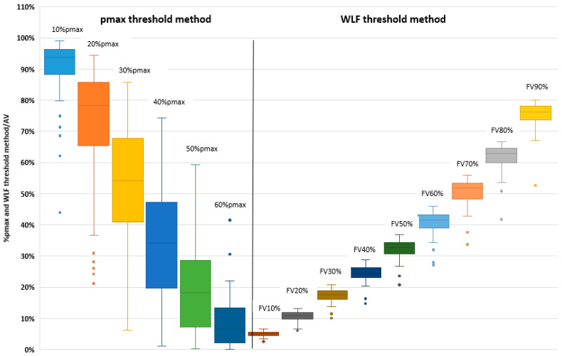
Material and method: Patients prospectively enrolled in the PEGASUS trial-a pilot study assessing the feasibility of lung functional avoidance using perfusion PET/CT imaging for lung stereotactic body radiotherapy (SBRT) of primary or secondary lesion-were analysed. Patients underwent lung perfusion MAA-68Ga PET/CT imaging and pulmonary function tests (PFTs) as part of pre-treatment evaluation. LFVs were delineated using two methods: the commonly used relative to the maximal pixel value threshold method (pmax threshold method, X%pmax volumes) and a new approach based on a relative to whole lung function threshold method (WLF threshold method, FVX% volumes) using a dedicated iterative algorithm. For both methods, LFVs were expressed in terms of % of the anatomical lung volume (AV) and of % of the total lung activity. Functional volumes were compared for patients with normal PFTs and pre-existing airway disease.
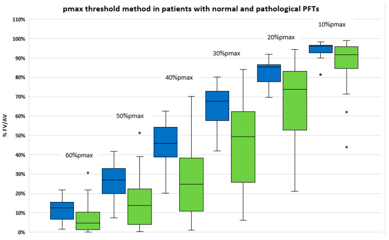
Results: 60 patients were analysed. Among the 48 patients who had PFTs, 31 (65%) had pre-existing lung disease. The pmax and WLF threshold methods clearly provided different functional volumes with a wide range of relative lung function for a given pmax volume, and conversely, a wide range of corresponding pmax values for a given WLF volume. The WLF threshold method provided more reliable and consistent volumes with much lower dispersion of LFVs as compared to the pmax method, especially in patients with normal PFTs.
Conclusions: We developed a relative to whole lung function threshold segmentation method to delineate lung functional volumes on perfusion PET/CT imaging. The automated algorithm allows for reproducible contouring. This new approach, relatively unaffected by the presence of hot spots, provides reliable and consistent functional volumes, and is clinically meaningful for clinicians.
Keywords: 68Ga-MAA-lung perfusion PET/CT; functional volumes; lung perfusion PET/CT; lung radiotherapy.
Conflict of interest statement
The authors declare no conflict of interest.
Figures

References
-
- Blanc-Béguin F., Hennebicq S., Robin P., Tripier R., Salaün P.-Y., Le Roux P.-Y. Radiopharmaceutical Labelling for Lung Ventilation/Perfusion PET/CT Imaging: A Review of Production and Optimization Processes for Clinical Use. Pharmaceuticals. 2022;15:518. doi: 10.3390/ph15050518. - DOI - PMC - PubMed
LinkOut - more resources
Full Text Sources
