. 2023 Mar 24;24(7):6125.
doi: 10.3390/ijms24076125.

Wds-Mediated H3K4me3 Modification Regulates Lipid Synthesis and Transport in Drosophila

Affiliations

Wds-Mediated H3K4me3 Modification Regulates Lipid Synthesis and Transport in Drosophila

Tujing Zhao et al. Int J Mol Sci. .

Abstract

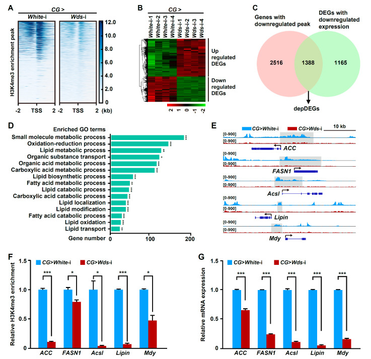
Lipid homeostasis is essential for insect growth and development. The complex of proteins associated with Set 1 (COMPASS)-catalyzed Histone 3 lysine 4 trimethylation (H3K4me3) epigenetically activates gene transcription and is involved in various biological processes, but the role and molecular mechanism of H3K4me3 modification in lipid homeostasis remains largely unknown. In the present study, we showed in Drosophila that fat body-specific knockdown of will die slowly (Wds) as one of the COMPASS complex components caused a decrease in lipid droplet (LD) size and triglyceride (TG) levels. Mechanistically, Wds-mediated H3K4me3 modification in the fat body targeted several lipogenic genes involved in lipid synthesis and the Lpp gene associated with lipid transport to promote their expressions; the transcription factor heat shock factor (Hsf) could interact with Wds to modulate H3K4me3 modification within the promoters of these targets; and fat body-specific knockdown of Hsf phenocopied the effects of Wds knockdown on lipid homeostasis in the fat body. Moreover, fat body-specific knockdown of Wds or Hsf reduced high-fat diet (HFD)-induced oversized LDs and high TG levels. Altogether, our study reveals that Wds-mediated H3K4me3 modification is required for lipid homeostasis during Drosophila development and provides novel insights into the epigenetic regulation of insect lipid metabolism.

Keywords: H3K4me3; Hsf; Wds; fat body; intestine; lipid synthesis; lipid transport.


Conflict of interest statement

The authors declare no conflict of interest. The funders had no role in the design of this study; in the collection, analyses, or interpretation of data; in the writing of the manuscript, or in the decision to publish the results.

Figures

Figure 1
Figure 1
Wds knockdown in the Drosophila fat body impaired lipid content. (AC) TRiP RNAi line (BS32952)-mediated Wds knockdown of Drosophila larvae using CG-Gal4 decreased the TG levels (n = 3, 10 larvae per group) of third instar larvae (A). This genetic manipulation for Wds was used hereafter unless otherwise indicated. Fat body-specific Wds knockdown decreased the LD size in the fat body of third instar larvae (B,C). Quantification of LDs size (C). Each point represents a single LD. BODIPY, green; DAPI, blue. Scale bar, 50 μm. (DF) VDRC RNAi line (V105371)-mediated Wds knockdown in the fat body using CG-Gal4 decreased TG levels (n = 3, 10 larvae per group) of third instar larvae (D) and reduced LD size (E,F) in the fat body of Drosophila third instar larvae. V60100 line was used as control. (GI) TRiP RNAi line (BS32952)-mediated Wds knockdown in the fat body using R4-Gal4 decreased TG levels (n = 3, 10 larvae per group) of third instar larvae (G) of Drosophila larvae and reduced LD size (H,I) in the fat body. Each point represents a single LD (H). BODIPY, green; DAPI, blue. Scale bar, 50 μm. Data are presented as the mean ± SE (error bars). For the significance: ** p < 0.01 and *** p < 0.001 versus the control.
Figure 2
Figure 2
Fat body-specific Wds knockdown reduced H3K4me3 deposition within the promoters of lipogenic genes and impaired their transcriptions. (A) ChIP-seq identified the change of H3K4me3 enrichment around genome-wide transcription start sites (TSSs) in the fat body of Drosophila third instar larvae following fat body-specific Wds knockdown. The heatmap of H3K4me3 ChIP peaks around the TSSs was constructed. (B) Hierarchical clustering of all differentially expressed genes (DEGs) induced by Wds knockdown in the fat body. (C) RNA-seq-based identification of differentially expressed genes with downregulated mRNA expression and downregulated H3K4me3 ChIP peaks (depDEGs) after fat body-specific Wds knockdown. (D) GO enrichment of depDEGs after fat body-specific Wds knockdown. (E) Fat body-specific Wds knockdown-caused changes in H3K4me3 ChIP peaks within the promoters of several lipogenic genes from the depDEGs, including ACC, FASN1, Acsl, Lipin, and Mdy. The representative peaks were highlighted in grey. Arrows indicate the direction of gene transcription. (F) ChIP-qPCR confirmation of fat body-specific Wds knockdown-caused decrease in H3K4me3 enrichment at lipogenic genes in the fat body of Drosophila third instar larvae. (G) RT-qPCR confirmation of fat body-specific Wds knockdown-caused downregulation in mRNA expression of lipogenic genes in the fat body (n = 3, 10 larvae per group). Data are presented as the mean ± SE (error bars). For the significance: * p < 0.05, ** p < 0.01, and *** p < 0.001 versus the control.
Figure 3
Figure 3
Fat body-specific knockdown of Wds downregulated Lpp transcription and impaired lipid transport. (A) Fat body-specific Wds knockdown-caused changes in H3K4me3 ChIP peaks within the promoter of the Lpp gene that encodes a lipoprotein mediating lipid transport from the intestine and was also included in the depDEGs. The representative peak was highlighted in grey. The arrow indicated transcription direction. (B) RNA-seq data of Wds knockdown-induced decrease in Lpp mRNA expression in the larval fat body. FPKM, fragments per kilo base of transcript per million mapped fragments. (C) ChIP-qPCR confirmation of fat body-specific Wds knockdown-caused decrease in H3K4me3 enrichment within the Lpp gene in the fat body of Drosophila third instar larvae. (D) Wds knockdown in the fat body downregulated Lpp transcription in the fat body (n = 3, 10 larvae per group). (E,F) Fat body-specific Wds knockdown caused elevated TG levels (n = 3, 20 larvae per group) (E) and lipid accumulation (F) in the third instar larvae intestine but decreased TG levels (n = 3, 60 larvae per group) in the hemolymph (G). BODIPY, green; DAPI, blue. Scale bar, 100 μm. Data are presented as the mean ± SE (error bars). For the significance: * p < 0.05 and *** p < 0.001 versus the control.
Figure 4
Figure 4
Hsf promoted lipid synthesis and transport by interacting with Wds to modulate H3K4me3 modification. (A) Schematic diagram for multiple approaches-based identification of the transcription factor Hsf that might interact with Wds, had potential binding motifs within the promoters of lipid homeostasis-related genes, and was involved in Drosophila lipid synthesis. (B,C) TRiP RNAi line (THU2458)-mediated Hsf knockdown in the Drosophila fat body reduced LD size (B) and decreased the TG levels (n = 3, 10 larvae per group) (C) of Drosophila third instar larvae. BODIPY, green; DAPI, blue. Scale bar, 50 μm. (DF) Fat body-specific Hsf knockdown caused elevated TG levels (n = 3, 20 larvae per group) (D) and lipid accumulation (E) in the intestine but decreased TG levels (n = 3, 60 larvae per group) in the hemolymph (F). BODIPY, green; DAPI, blue. Scale bar, 100 μm (E). (G) Co-IP confirmation of the interaction between Flag-tagged Hsf and HA-tagged Wds that were transiently expressed in Drosophila S2 cells. The antibodies against the HA tag or Flag tag were used. Tubulin was used as the loading control. (H,I) Fat body-specific Hsf knockdown reduced H3K4me3 enrichment within promoters of lipogenic genes and Lpp (H) as well as decreased the transcriptions of these lipid homeostasis-related genes (I) in the fat body. Data are presented as the mean ± SE (error bars). For the significance: * p < 0.05, ** p < 0.01, and *** p < 0.001 versus the control.
Figure 5
Figure 5
HFD increased the H3K4me3 levels to regulate lipid synthesis. (A) HFD feeding increased H3K4me3 levels within the promoters of lipogenic genes in the Drosophila fat body. (B) RT-qPCR confirmation of HFD-mediated upregulation in Wds mRNA expression in the fat body (n = 3, 10 larvae per group). (C) Western blotting confirmed the HFD-mediated increase in protein expression of Wds in the fat body. (D) RT-qPCR confirmation of HFD-mediated upregulation of the Hsf expression in the fat body (n = 3, 10 larvae per group). (EG) Fat body-specific Wds knockdown could rescue HFD-induced defects in TG levels (n = 3, 10 larvae per group) of Drosophila larvae and LD size (F,G) in the fat body. Quantification of LD size in (G). Each point is a single LD. BODIPY, green; DAPI, blue. Scale bar, 50 μm. (HJ) Fat body-specific Hsf knockdown could rescue HFD-induced defects in TG levels (n = 3, 10 larvae per group) (H) of Drosophila larvae and LD size in the fat body. Each point is a single LD. BODIPY, green; DAPI, blue. Scale bar, 50 μm. ND, normal diet; HFD, high-fat diet. Data are presented as the mean ± SE (error bars). For the significance: * p < 0.05, ** p < 0.01, and *** p < 0.001 versus the control.

Similar articles

References

    1. Olzmann J.A., Carvalho P. Dynamics and functions of lipid droplets. Nat. Rev. Mol. Cell. Biol. 2019;20:137–155. doi: 10.1038/s41580-018-0085-z. - DOI - PMC - PubMed
    1. Walther T.C., Farese R.V., Jr. Lipid droplets and cellular lipid metabolism. Annu. Rev. Biochem. 2012;81:687–714. doi: 10.1146/annurev-biochem-061009-102430. - DOI - PMC - PubMed
    1. Welte M.A. As the fat flies: The dynamic lipid droplets of Drosophila embryos. Biochim. Biophys. Acta. 2015;1851:1156–1185. doi: 10.1016/j.bbalip.2015.04.002. - DOI - PMC - PubMed
    1. Nelliot A., Bond N., Hoshizaki D.K. Fat-body remodeling in Drosophila melanogaster. Genesis. 2006;44:396–400. doi: 10.1002/dvg.20229. - DOI - PubMed
    1. Li S., Yu X., Feng Q. Fat body biology in the last decade. Annu. Rev. Entomol. 2019;64:315–333. doi: 10.1146/annurev-ento-011118-112007. - DOI - PubMed

LinkOut - more resources