. 2023 Mar 24;24(7):6144.
doi: 10.3390/ijms24076144.

Low P-Selectin Glycoprotein Ligand-1 Expression in Neutrophils Associates with Disease Activity and Deregulated NET Formation in Systemic Lupus Erythematosus

Affiliations

Low P-Selectin Glycoprotein Ligand-1 Expression in Neutrophils Associates with Disease Activity and Deregulated NET Formation in Systemic Lupus Erythematosus

Antonio Muñoz-Callejas et al. Int J Mol Sci. .

Abstract

Systemic Lupus Erythematosus (SLE) is an autoimmune disease characterized by the generation of anti-DNA autoantibodies due to exposure of immune cells to excessive amounts of extracellular DNA. Lack of P-selectin in mice induces the development of a lupus-like syndrome and patients with cutaneous lupus have reduced P-selectin expression in skin vessels. Using flow cytometry we analyzed in healthy donors and patients the expression of P-selectin Glycoprotein Ligand-1 (PSGL-1) in circulating neutrophils and the implication of PSGL-1/P-selectin interaction in neutrophil extracellular traps (NETs) generation. We found a statistical significance that neutrophils from active SLE patients have a reduced expression of PSGL-1 and low levels of PSGL-1 in neutrophils from SLE patients associated with the presence of anti-dsDNA antibodies, clinical lung involvement, Raynaud's phenomenon, and positive lupus anticoagulant. PSGL-1 is present along the DNA in the NET. In healthy donors, neutrophil interaction with immobilized P-selectin triggers Syk activation, increases the NETs percentage and reduces the amount of DNA extruded in the NETs. In active SLE patients, neutrophil interaction with P-selectin does not activate Syk or reduce the amount of DNA extruded in the NETs, that might contribute to increase the extracellular level of DNA and hence, to disease pathogenesis.

Keywords: NETs; P-selectin; PSGL-1; SLE pathogenesis; neutrophils.


Conflict of interest statement

The authors declare no conflict of interest.

Figures

Figure 1
Figure 1
PSGL-1 expression in circulating neutrophils from SLE patients and healthy donors. Expression of PSGL-1 was analyzed by flow cytometry. (a) Total (left panel) and membrane (right panel) expression of PSGL-1 in neutrophils from healthy donors (HD, n = 41), inactive SLE patients (iSLE, n = 33), and active SLE patients (aSLE, n = 14). (be) Association of membrane and total PSGL-1 expression in neutrophils from SLE patients with the presence of different clinical characteristics: (b) presence of anti-dsDNA autoantibodies in serum (n = 47), (c) lupus anticoagulant positivity (n = 25), (d) lung disease involvement (n = 47), and (e) presence of Raynaud´s phenomenon (n = 47). Data represent mean fluorescence intensity (MFI) ± SEM. Differences were analyzed by Student’s t-test. * p ≤ 0.05; ** p ≤ 0.01. HD: healthy donors. SLE: Systemic Lupus Erythematosus. iSLE: inactive SLE. aSLE: active SLE.
Figure 2
Figure 2
PSGL-1 localization in neutrophils and NETs. Representative microphotographs of neutrophils isolated from blood of HD, after 1 h incubation on BSA and labeled with anti-PSGL-1 monoclonal antibody plus anti-mouse-Alexa Fluor555 (red) secondary antibody, anti-neutrophil elastase rabbit polyclonal antibody plus anti-rabbit-Alexa Fluor 647 (green) secondary antibody and DAPI to visualize DNA (cyan). (a) Resting neutrophils. (b,c) Spontaneous NETs. (d) Scatterplot representing Mander’s coefficient for PSGL-1 and NE co-localization in resting neutrophils (0.74 ± 0.21) and NETs (0.67 ± 0.21). Scale bar = 2 µm. All images were obtained by confocal microscopy with 63× oil objective and a 5× digital zoom. Arrowheads indicate cytoplasmic granules. Arrows indicate the neutrophil membrane. NETs: Neutrophil extracellular traps. NE: Neutrophil elastase. PSGL-1: P-selectin glycoprotein ligand-1.
Figure 3
Figure 3
Susceptibility to NET generation of neutrophils from SLE patients and healthy donors in basal experimental conditions. Quantification of NETs generated by neutrophils from HD, iSLE patients and aSLE patients. (a) Representative fields of DAPI/NE stained unstimulated neutrophils from HD, iSLE and aSLE obtained by confocal microscopy with a 63x objective. Scale bar = 10–50 µm. Yellow arrowheads indicate NETs. Yellow asterisks indicate NETosing cells amplified below the image. (b) Percentage of NETs quantified by confocal microscopy. (c,d) MFI of Sytox Green in (c) Sytox Green/NE-APC and (d) Sytox Green/MPO-PE double positive events, respectively. Statistical analysis was performed using unpaired Student’s t-test and Mann-Whitney test. * p ≤ 0.05; ** p ≤ 0.01, *** p ≤ 0.001. Data show mean fluorescence intensity (MFI) ± SEM or percentage ± SEM. HD (n = 6–12), iSLE (n = 4–7), aSLE (n = 3–8). HD: healthy donors. SLE: Systemic Lupus Erythematosus. iSLE: inactive SLE. aSLE: active SLE. NETs: Neutrophil extracellular traps. NE: Neutrophil Elastase. MPO: Myeloperoxidase. SLE = Total population of patients included.
Figure 4
Figure 4
Interaction of HD neutrophils with P-selectin regulates NET generation. Peripheral neutrophils isolated from healthy donors were incubated in rolling-like conditions. (a) Representative confocal microphotographs taken with 63× and 63× plus 5× digital zoom of DAPI/NE staining neutrophils in rolling-like experimental conditions on BSA or BSA+P-selectin. Scale bar = 10–50 µm. Yellow arrowheads indicate NETs. Yellow asterisks indicate NETosing cells amplified below the image. (b) Percentage of NETs quantified by confocal microscopy. (c,d) Percentage of NETosing cells in (c) Sytox Green/NE-APC and (d) Sytox Green/MPO-PE double positive events, respectively. (e) Extracellular Sytox Green MFI analyzed by fluorometry at 485 nm. (f,g) Sytox Green MFI fold induction in (f) Sytox Green/NE-APC and (g) Sytox Green/MPO-PE double positive events, respectively. Data are presented as MFI fold induction or percentage ± SEM. Statistical analysis was performed using paired Student’s t-test and Wilcoxon test. * p ≤ 0.05; ** p ≤ 0.01; n = 6–12. NETs: Neutrophil extracellular traps. HD: healthy donors. NE: Neutrophil elastase. MPO: Myeloperoxidase.
Figure 5
Figure 5
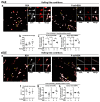
Deregulated NET generation by neutrophils from SLE patients. Neutrophils from inactive (iSLE) and active (aSLE) patients were incubated in rolling-like conditions in the presence of BSA or BSA+P-selectin and NETs were quantified by confocal microscopy and by flow cytometry. (ad) iSLE patients: (a) Representative confocal microphotographs of DAPI/NE stained neutrophils. (b) Quantification of NETs by confocal microscopy. (c,d) MFI fold induction of Sytox Green in (c) Sytox Green/NE double positive events and (d) Sytox Green/MPO double positive events. (eh) aSLE patients: (e) Representative confocal microphotographs of DAPI/NE stained neutrophils. (f) Quantification of NETs by confocal microscopy. (g,h) MFI fold induction of Sytox Green in (g) Sytox Green/NE double positive events and (h) Sytox Green/MPO double positive events. Confocal images were taken with 63× and 63× plus 5× digital zoom. Scale bar = 10–50 µm. Yellow arrowheads indicate NETs. Yellow numbers 1 and 2 indicate NETosing cells amplified next to the 63× image. Data are presented as mean fluorescence intensity (MFI) fold induction or percentage ± SEM. Statistical analysis was performed using paired Student’s t-test and Wilcoxon test. * p ≤ 0.05, ** p ≤ 0.01. iSLE patients (n = 4–7), aSLE patients (n = 6–8). SLE: Systemic Lupus Erythematosus. iSLE: inactive SLE. aSLE: active SLE. NETs: Neutrophil extracellular traps.
Figure 6
Figure 6
Expression of L-selectin and pSyk in neutrophils from healthy donors and SLE patients in experimental conditions. (a,d) Gating strategy for identification of (a) L-selectin, and (d) pSyk and pSykHigh populations: Singlets were identified by FSC-H and FSC-A criteria. Neutrophil populations were identified in the singlets gate by SSC-A/FSC-A criteria. Gates for positive cells were identified by using isotype controls. (b,c) L-selectin expression in basal experimental conditions: (b) Percentage of cells expressing L-selectin, and (c) MFI of L-selectin. (e,f) Percentage of neutrophils expressing (e) pSyk or (f) pSykHigh in basal experimental conditions. (gj) pSyk and pSykHigh expression in HD neutrophils in rolling-like conditions: (g) MFI fold induction of pSyk, (h) Percentage of pSyk expressing neutrophils, (i) MFI of pSykHigh, and (j) Percentage of pSykHigh expressing neutrophils. (kn) pSyk and pSykHigh expression in iSLE neutrophils in rolling-like conditions: (k) MFI fold induction of pSyk, (l) Percentage of pSyk expressing neutrophils, (m) MFI fold induction of pSykHigh, and (n) Percentage of pSykHigh expressing neutrophils. (or) pSyk and pSykHigh expression in aSLE neutrophils in rolling-like conditions: (o) MFI fold induction of pSyk, (p) Percentage of pSyk expressing cells, (q) MFI fold induction of pSykHigh, and (r) Percentage of pSykHigh expressing neutrophils. Data are presented as MFI fold induction or percentage ± SEM. Statistical analysis was performed using paired Student’s t-test and Wilcoxon test. * p ≤ 0.05; ** p ≤ 0.01, **** p ≤ 0.0001. HD (n = 7–12). iSLE patients (n = 7), aSLE patients (n = 7–8). MFI: Mean fluorescence intensity. HD: Healthy donor. SLE: Systemic Lupus Erythematosus. iSLE: inactive SLE. aSLE: active SLE. SLE: Total population of patients included. Open circles: basal conditions. Open diamonds: rolling-like on BSA. Black diamonds: rollink-like on P-selectin.
Figure 7
Figure 7
Rolling-like assays in neutrophils isolated from peripheral blood. Created with Biorender.com.

References

    1. Prame Kumar K., Nicholls A.J., Wong C.H.Y. Partners in crime: Neutrophils and monocytes/macrophages in inflammation and disease. Cell Tissue Res. 2018;371:551–565. doi: 10.1007/s00441-017-2753-2. - DOI - PMC - PubMed
    1. Brinkmann V., Reichard U., Goosmann C., Fauler B., Uhlemann Y., Weiss D.S., Weinrauch Y., Zychlinsky A. Neutrophil extracellular traps kill bacteria. Science. 2004;303:1532–1535. doi: 10.1126/science.1092385. - DOI - PubMed
    1. Dorner T., Furie R. Novel paradigms in systemic lupus erythematosus. Lancet. 2019;393:2344–2358. doi: 10.1016/S0140-6736(19)30546-X. - DOI - PubMed
    1. Fortuna G., Brennan M.T. Systemic lupus erythematosus: Epidemiology, pathophysiology, manifestations, and management. Dent. Clin. N. Am. 2013;57:631–655. doi: 10.1016/j.cden.2013.06.003. - DOI - PubMed
    1. Hakkim A., Furnrohr B.G., Amann K., Laube B., Abed U.A., Brinkmann V., Herrmann M., Voll R.E., Zychlinsky A. Impairment of neutrophil extracellular trap degradation is associated with lupus nephritis. Proc. Natl. Acad. Sci. USA. 2010;107:9813–9818. doi: 10.1073/pnas.0909927107. - DOI - PMC - PubMed