

Characterization of the Protein Corona of Three Chairside Hemoderivatives on Melt Electrowritten Polycaprolactone Scaffolds
- PMID: 37047135
- PMCID: PMC10094244
- DOI: 10.3390/ijms24076162
Characterization of the Protein Corona of Three Chairside Hemoderivatives on Melt Electrowritten Polycaprolactone Scaffolds
Abstract
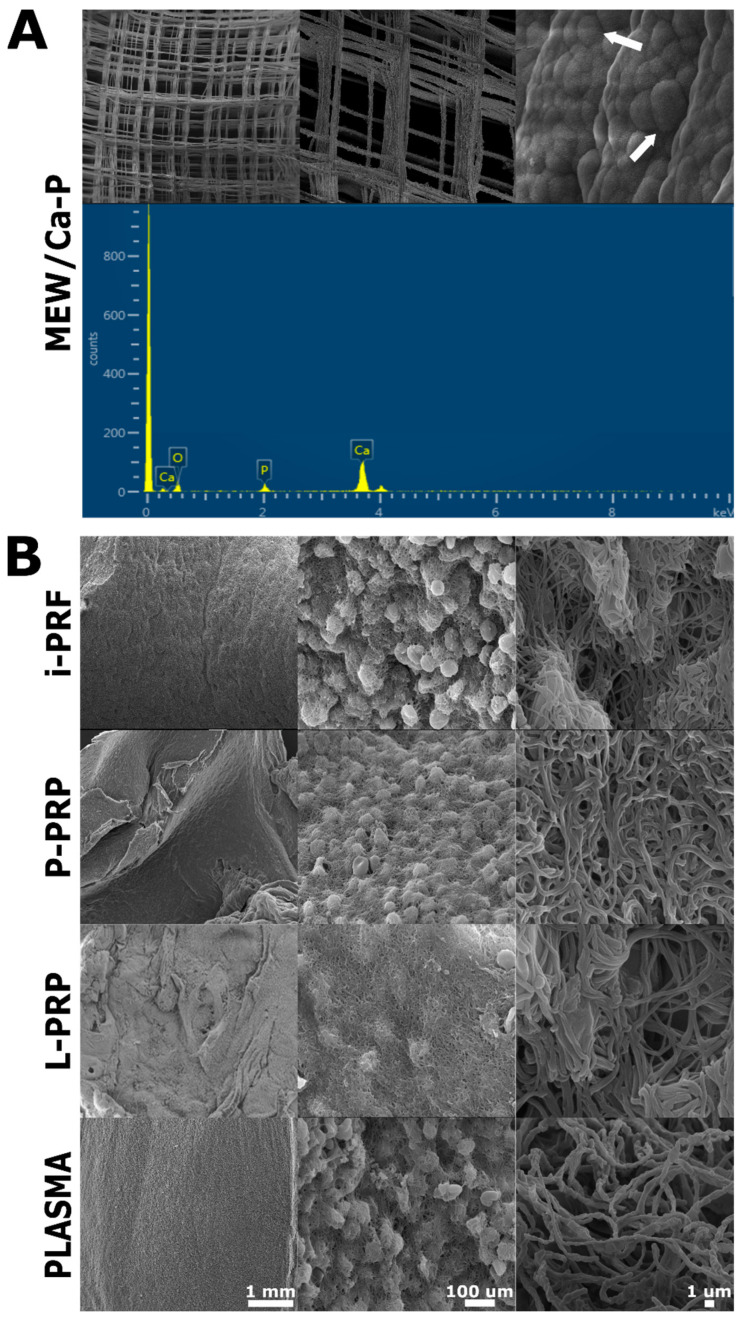
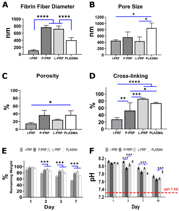
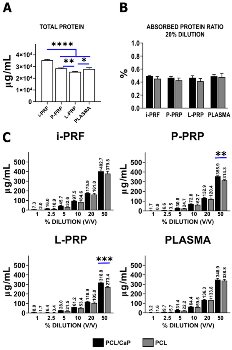
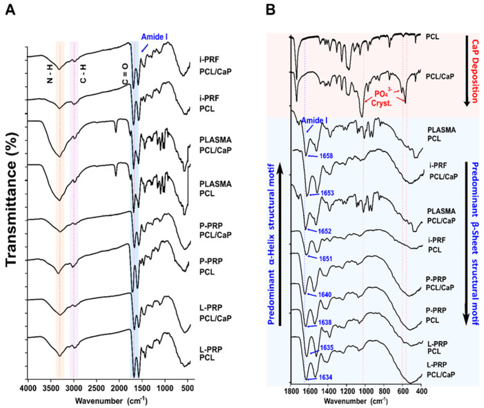
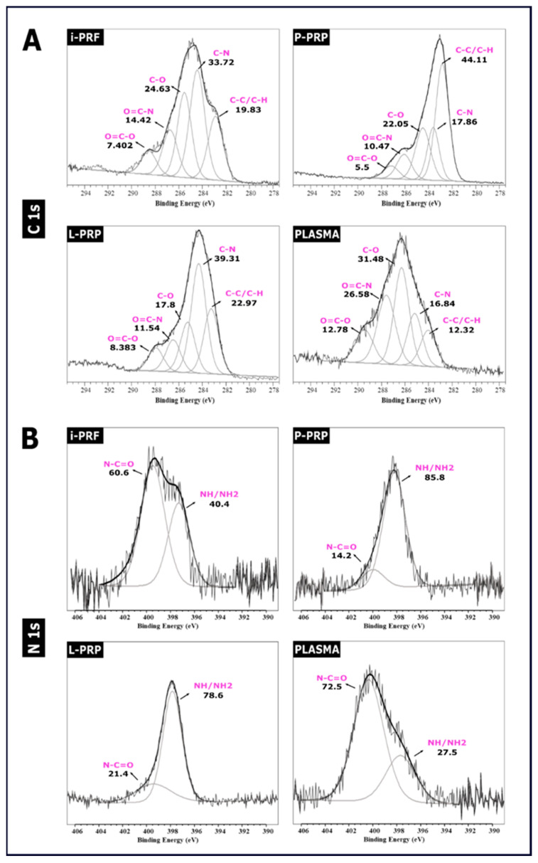
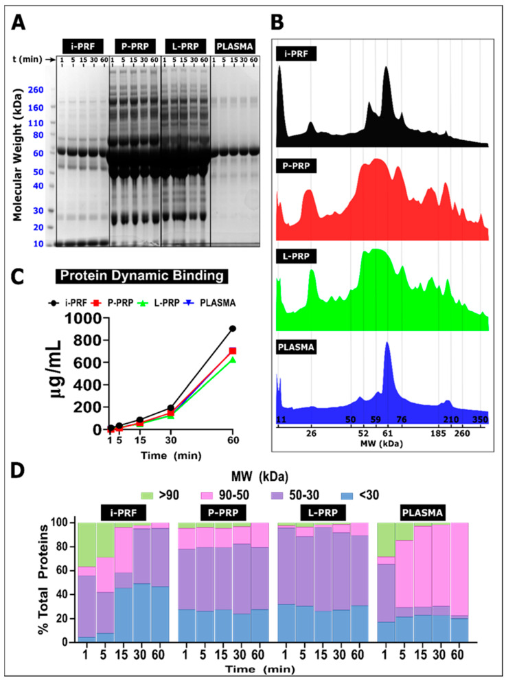
In tissue engineering, the relationship between a biomaterial surface and the host's immune response during wound healing is crucial for tissue regeneration. Despite hemoderivative functionalization of biomaterials becoming a common tissue-engineering strategy for enhanced regeneration, the characteristics of the protein-biomaterial interface have not been fully elucidated. This study characterized the interface formed by the adsorbed proteins from various hemoderivatives with pristine and calcium phosphate (CaP)-coated polycaprolactone (PCL) melt electrowritten scaffolds. PCL scaffolds were fabricated by using melt electrospinning writing (MEW). Three hemoderivatives (pure platelet-rich plasma (P-PRP), leucocyte platelet-rich plasma (L-PRP) and injectable platelet-rich fibrin (i-PRF)) and total blood PLASMA (control) were prepared from ovine blood. Hemoderivatives were characterized via SEM/EDX, cross-linking assay, weight loss, pH and protein quantification. The interface between PCL/CaP and hemoderivative was examined via FTIR, XPS and electrophoresis. i-PRF/PCL-CaP (1653 cm-1), PLASMA/PCL-CaP (1652 cm-1) and i-PRF/PCL (1651 cm-1) demonstrated a strong signal at the Amide I region. PLASMA and i-PRF presented similar N1s spectra, with most of the nitrogen involved in N-C=O bonds (≈400 eV). i-PRF resulted in higher adsorption of low molecular weight (LMW) proteins at 60 min, while PLASMA exhibited the lowest adsorption. L-PRP and P-PRP had a similar pattern of protein adsorption. The characteristics of biomaterial interfaces can be customized, thus creating a specific hemoderivative-defined layer on the PCL surface. i-PRF demonstrated a predominant adsorption of LMW proteins. Further investigation of hemoderivative functionalized biomaterials is required to identify the differential protein corona composition, and the resultant immune response and regenerative capacity.
Keywords: hemoderivative; polycaprolactone; protein corona; regeneration.
Conflict of interest statement
The authors declare no conflict of interest.
Figures
Similar articles
-
Mesenchymal stem cells in PRP and PRF containing poly(3-caprolactone)/gelatin Scaffold: a comparative in-vitro study.Cell Tissue Bank. 2024 Jun;25(2):559-570. doi: 10.1007/s10561-023-10116-x. Epub 2024 Feb 16. Cell Tissue Bank. 2024. PMID: 38363442
-
Systematic Comparison of the Effect of Four Clinical-Grade Platelet Rich Hemoderivatives on Osteoblast Behaviour.Int J Mol Sci. 2019 Dec 11;20(24):6243. doi: 10.3390/ijms20246243. Int J Mol Sci. 2019. PMID: 31835696 Free PMC article.
-
Injectable-platelet rich fibrin using the low speed centrifugation concept improves cartilage regeneration when compared to platelet-rich plasma.Platelets. 2019;30(2):213-221. doi: 10.1080/09537104.2017.1401058. Epub 2017 Dec 14. Platelets. 2019. PMID: 29240523
-
In search of a consensus terminology in the field of platelet concentrates for surgical use: platelet-rich plasma (PRP), platelet-rich fibrin (PRF), fibrin gel polymerization and leukocytes.Curr Pharm Biotechnol. 2012 Jun;13(7):1131-7. doi: 10.2174/138920112800624328. Curr Pharm Biotechnol. 2012. PMID: 21740379 Review.
-
Classification of platelet concentrates: from pure platelet-rich plasma (P-PRP) to leucocyte- and platelet-rich fibrin (L-PRF).Trends Biotechnol. 2009 Mar;27(3):158-67. doi: 10.1016/j.tibtech.2008.11.009. Epub 2009 Jan 31. Trends Biotechnol. 2009. PMID: 19187989 Review.
Cited by
-
Navigating the combinations of platelet-rich fibrin with biomaterials used in maxillofacial surgery.Front Bioeng Biotechnol. 2024 Oct 7;12:1465019. doi: 10.3389/fbioe.2024.1465019. eCollection 2024. Front Bioeng Biotechnol. 2024. PMID: 39434715 Free PMC article. Review.
-
Impact of autologous platelet concentrates on the osseointegration of dental implants.Periodontol 2000. 2025 Feb;97(1):271-286. doi: 10.1111/prd.12563. Epub 2024 Apr 22. Periodontol 2000. 2025. PMID: 38647020 Free PMC article. Review.
References
MeSH terms
Substances
LinkOut - more resources
Full Text Sources
Research Materials
Miscellaneous
