Review
. 2023 Mar 24;24(7):6187.
doi: 10.3390/ijms24076187.

Allosteric Regulation of G-Protein-Coupled Receptors: From Diversity of Molecular Mechanisms to Multiple Allosteric Sites and Their Ligands

Affiliations
Review

Allosteric Regulation of G-Protein-Coupled Receptors: From Diversity of Molecular Mechanisms to Multiple Allosteric Sites and Their Ligands

Alexander O Shpakov. Int J Mol Sci. .

Abstract

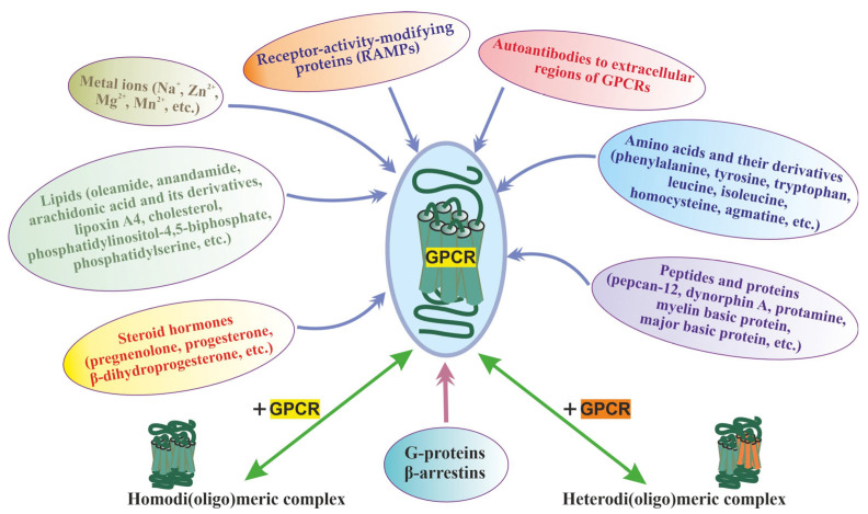
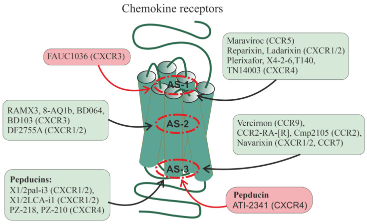
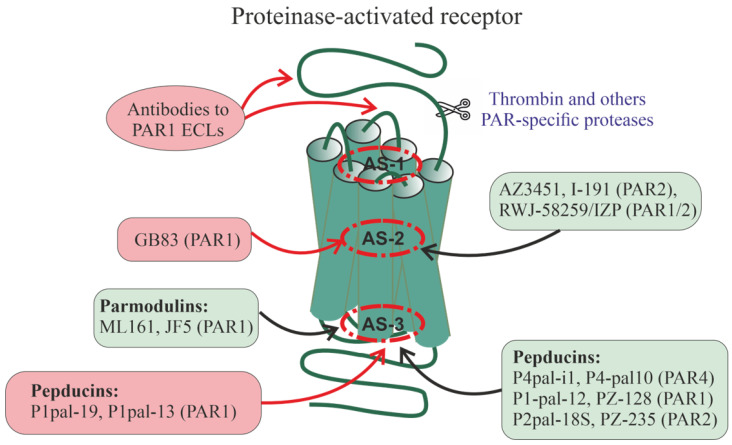
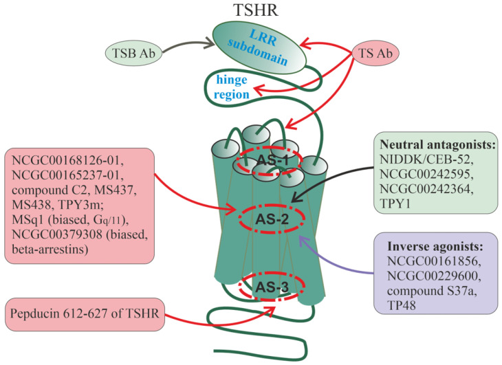
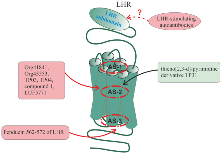
Allosteric regulation is critical for the functioning of G protein-coupled receptors (GPCRs) and their signaling pathways. Endogenous allosteric regulators of GPCRs are simple ions, various biomolecules, and protein components of GPCR signaling (G proteins and β-arrestins). The stability and functional activity of GPCR complexes is also due to multicenter allosteric interactions between protomers. The complexity of allosteric effects caused by numerous regulators differing in structure, availability, and mechanisms of action predetermines the multiplicity and different topology of allosteric sites in GPCRs. These sites can be localized in extracellular loops; inside the transmembrane tunnel and in its upper and lower vestibules; in cytoplasmic loops; and on the outer, membrane-contacting surface of the transmembrane domain. They are involved in the regulation of basal and orthosteric agonist-stimulated receptor activity, biased agonism, GPCR-complex formation, and endocytosis. They are targets for a large number of synthetic allosteric regulators and modulators, including those constructed using molecular docking. The review is devoted to the principles and mechanisms of GPCRs allosteric regulation, the multiplicity of allosteric sites and their topology, and the endogenous and synthetic allosteric regulators, including autoantibodies and pepducins. The allosteric regulation of chemokine receptors, proteinase-activated receptors, thyroid-stimulating and luteinizing hormone receptors, and beta-adrenergic receptors are described in more detail.

Keywords: G protein-coupled receptor; allosteric modulator; allosteric site; autoantibody; chemokine receptor; heterotrimeric G protein; luteinizing hormone receptor; pepducin; proteinase-activated receptor; thyroid-stimulating hormone receptor.


Conflict of interest statement

The author declares no conflict of interest.

Figures

Figure 1
Figure 1
The main group of allosteric regulators of GPCRs, including the homo- and heterodimerization, along with the main classes of allosteric regulators (ions, lipids, amino acids, peptides, proteins, steroid hormones, autoantibodies to extracellular regions of GPCRs), the heterotrimeric G proteins, β-arrestins, and RAMPs are shown, which form complexes with GPCRs during signal transduction acting allosterically on their functional activity. The allosteric effect of homo- and heterodi(oligo)merization of the receptors has also been shown, as demonstrated for class C GPCRs and some representatives of the class.
Figure 2
Figure 2
Allosteric regulation of chemokine receptors. There are data on the allosteric regulation of a significant number of chemokine receptors, including CXCR1, CXCR2, CXCR3, CXCR4, CCR2, CCR5, CCR7, and CCR9. Allosteric sites in them can be located at different loci of the TMD: in the upper (outwardly oriented) portion of the transmembrane tunnel (interfaces including the membrane-proximal segments of ECLs and extracellular segments of TMs, as well as the outer vestibule of the transmembrane channel) (designated as AS-1), in the central part of the 7TM bundle (AS-2), and in the lower (oriented to the cytoplasm) portion of the transmembrane tunnel (interfaces, including the membrane-proximal segments of ICLs and the cytoplasmic endings of TMs) (AS-3). In each of these loci, there are several cavities in which allosteric sites can be located, both overlapping and spatially separated. Along with this, allosteric sites can be located in hydrophilic loops being targets for autoantibodies and synthetic pepducins. Endogenous allosteric regulators of the chemokine receptors are sodium (NAM) and zinc (PAM) ions, as well as cholesterol and membrane phospholipids (mainly with PAM activity). Synthetic small allosteric regulators interact with allosteric sites located at all three loci, AS-1, AS-2, and AS-3, while pepducins, lipidated derivatives of peptides corresponding to ICLs, are transported across the plasma membrane and interact with intracellular sites. Figure 2 shows the following small regulators interacting with the AS-1 locus: maraviroc (CCR5-selective NAM), reparixin, ladarixin and their analogs (CXCR1/CXCR2-selective NAMs and/or non-competitive allosteric antagonists), plerixafor (allosteric CXCR4-inhibitor), FAUC1036 (biased allosteric CXCR3-agonist), T140 and its analog TN14003 (biased allosteric CXCR4-antagonists), and the peptide X4-2-6 (TM2/ECL1 of CXCR4; allosteric CXCR4-antagonist). The following ligands bind specifically to the AS-2 locus: RAMX3 and its biased analogs 8-azaquinazolinone (8-AQ) derivative 1b, BD064, and BD103 (CXCR3-selective NAMs), DF2755A (noncompetitive allosteric CXCR1/CXCR2-antagonist). The following ligands bind specifically to the AS-3 locus: vercirnon and its analogs (allosteric CCR9-antagonists), navarixin (allosteric antagonist of CXCR1, CXCR2 and CCR7), CCR2-RA-[R] and Cmp2105 (allosteric CCR2-antagonists and/or NAMs). Pepducins, such as N-palmitoylated peptides X1/2pal-i3 (ICL3 of CXCR1/CXCR2; CXCR1/CXCR2-selective NAM), PZ-218 (ICL1 of CXCR4; CXCR4-selective NAM), and PZ-210 (ICL3 of CXCR4; CXCR4-selective NAM), as well as lithocholic acid-modified X1/2LCA-i1 (ICL1 CXCR1/CXCR2; allosteric CXCR1/CXCR2-inhibitor and/or NAM) and ATI-2341 (ICL1 of CXCR4; CXCR4-selective PAM) interact with intracellular sites involved in functional coupling with G proteins and β-arrestins. Antibodies against extracellular regions of chemokine receptors are able to interact with ECLs and the extracellular N-terminal domain, but their effects remain poorly understood. Allosteric regulators that increase receptor activity (full agonists) are placed in red squares, while allosteric regulators that decrease receptor activity (antagonists and NAMs) are placed in green squares.
Figure 3
Figure 3
Allosteric regulation of proteinase-activated receptors. There is now evidence of allosteric regulation and localization of allosteric sites for PAR1, PAR2, and PAR4. As in the case of chemokine receptors (see Figure 2), allosteric sites in PARs can be located at different TMD loci (AS-1, AS-2, and AS-3) in the ECLs where they are available for interaction with autoantibodies, as well as in the ICLs where they are targets for pepducins. The small compounds, such as GB83 (PAR1-selective PAM), AZ3451 (allosteric PAR2-inhibitor), I-191 (PAR2-selective NAM), and cross-linked heterobivalent allosteric PAR1/PAR2 inhibitor, RWJ-58259/imidazopyridazine (IZP) derivative), interact with allosteric sites located in the upper and central parts of the TM7 bundle. The low-molecular-weight parmodulins, including the PAR1-allosteric inhibitors ML161 (PM2) and JF5, as well as pepducins, interact with intracellular sites, including the AS-3 locus. The following pepducins have been obtained and have specific activity for PAR1, PAR2, and PAR4: Palm-SGRRYGHALR (P4pal-10) (ICL3 of PAR4, allosteric PAR4-antagonist and NAM and allosteric antagonist for the non-cognate receptors PAR1, FPR2, and FFAR2), Palm-ATGAPRLPST (P4pal-i1) (ICL1 of PAR4, allosteric PAR4-inhibitor), Palm-RCLSSSAVANRS (P1pal-12), Palm-RSLSSSAVANRS (P1pal-12S) and its analog P1pal-7 (PZ-128) (ICL3 of PAR1, allosteric PAR1-antagonists), Pal-RCLSSSAVANRSKKSRALF (P1pal-19), Palm-AVANRSKKSRALF (P1pal-13) (ICL3 of PAR1, full allosteric PAR1-agonists), and Palm-RSSAMDENSEKKRKSAIK270−287 (P2pal-18S) and its analog PZ-235 (ICL3 of PAR2, allosteric PAR1/2-antagonists). Data on autoantibodies to PARs are fragmentary, but antibodies to extracellular regions of PAR1 in patients with systemic sclerosis have been shown to function as PAR1-specific allosteric full agonists and/or PAMs. Many of the effects of allosteric regulators are due to their effect on the stability of heterodimeric PAR1/PAR2 complexes, as shown for the heterobivalent PAR1/PAR2-inhibitor and pepducins P1pal-12S and P2pal-18S. Allosteric regulators that increase receptor activity (full agonists and PAMs) are placed in red squares, while allosteric regulators that decrease receptor activity (antagonists and NAMs) are placed in green squares.
Figure 4
Figure 4
Allosteric regulators of thyroid-stimulating hormone receptor. The TSHR belongs to a family of class A GPCRs having a large extracellular domain in which an orthosteric site is located and which is a target for autoantibodies. Most of the small regulators of TSHR bind to allosteric sites located in the cavity of the AS-2 locus or slightly higher in the lower portions of the AS-1 locus. The following low-molecular-weight compounds specifically interact with allosteric sites in the AS-2 locus: compound C2, NCGC00168126-01 and its analog NCGC00165237-01 (full allosteric TSHR-agonists), MS437, MS438 (biased allosteric TSHR-agonists, did not affect Gi-proteins), TPY3m (biased allosteric TSHR-agonist, mainly activates Gs-proteins), MSq1 (biased allosteric TSHR-agonist, mainly activates Gq/11-proteins), NCGC00379308 (biased PAM, predominantly activates β-arrestins), TPY1, NIDDK/CEB-52 and its analogs NCGC00242595 and NCGC00242364 (allosteric TSHR-antagonists), NCGC00161856, NCGC00229600, compound S37a, and TP48 (allosteric inverse TSHR-agonists). Pepducin 612–627(Palm) (ICL3 of TSHR) interacts with the intracellular allosteric site and functions as an allosteric TSHR-agonist. Stimulating TSHR autoantibodies (TSAb) interact with the LRR subdomain, hinge region, and ECLs. Blocking TSHR autoantibodies (TSBAb) mainly interact with the N-terminal and central portions of the LRR subdomain. Allosteric regulators that increase receptor activity (full agonists and PAMs) are placed in red squares, allosteric regulators with neutral antagonist activity are placed in green squares, while allosteric regulators with inverse agonist activity are placed in blue squares.
Figure 5
Figure 5
Allosteric regulators of the luteinizing hormone receptor. Like TSHR, LHR has a large extracellular domain in which an orthosteric site is located, and the small LHR regulators bind to allosteric sites located in the cavity of the AS-2 locus. The following low-molecular-weight compounds specifically interact with allosteric sites in the AS-2 locus: thieno[2,3-d]-pyrimidine derivatives Org41841, Org43553, TP03, TP04 and their analogs (allosteric LHR-agonists or ago-PAMs, predominantly activate Gs-proteins), the derivatives of 1,3,5-pyrazole (compound 1) and terphenyl (LUF5771) (allosteric LHR-agonists), and thieno[2,3-d]-pyrimidine derivative TP31 (allosteric LHR-antagonists). Pepducin NKDTKIAKK-Nle-A(562–572)-K(Palm)A (ICL3 of LHR) interacts with the intracellular allosteric site and functions as an allosteric LHR agonist. Autoantibodies to the extracellular regions of LHR have been suggested but have not been characterized to date. Allosteric regulators that increase receptor activity (full agonists and ago-PAMs) are placed in red squares, while allosteric regulators with neutral antagonist activity are placed in green squares.

Similar articles

Cited by

References

    1. Brown N.A., Schrevens S., van Dijck P., Goldman G.H. Fungal G-Protein-Coupled Receptors: Mediators of Pathogenesis and Targets for Disease Control. Nat. MicroBiol. 2018;3:402–414. doi: 10.1038/s41564-018-0127-5. - DOI - PubMed
    1. Shpakov A.O., Pertseva M.N. Signaling Systems of Lower Eukaryotes and Their Evolution. Int. Rev. Cell Mol. Biol. 2008;269:151–282. doi: 10.1016/S1937-6448(08)01004-6. - DOI - PubMed
    1. Krishnan A., Almén M.S., Fredriksson R., Schiöth H.B. The origin of GPCRs: Identification of mammalian like Rhodopsin, Adhesion, Glutamate and Frizzled GPCRs in fungi. PLoS ONE. 2012;7:e29817. doi: 10.1371/journal.pone.0029817. - DOI - PMC - PubMed
    1. Liu Y., Wang X., Dong D., Guo L., Dong X., Leng J., Zhao B., Guo Y.-D., Zhang N. Research Advances in Heterotrimeric G-Protein α Subunits and Uncanonical G-Protein Coupled Receptors in Plants. Int. J. Mol. Sci. 2021;22:8678. doi: 10.3390/ijms22168678. - DOI - PMC - PubMed
    1. Reboul J., Ewbank J.J. GPCRs in Invertebrate Innate Immunity. Biochem. Pharmacol. 2016;114:82–87. doi: 10.1016/j.bcp.2016.05.015. - DOI - PubMed

MeSH terms

LinkOut - more resources