. 2023 Mar 25;24(7):6210.
doi: 10.3390/ijms24076210.

Amniotic Membrane Restores Chronic Wound Features to Normal in a Keratinocyte TGF-β-Chronified Cell Model

Affiliations

Amniotic Membrane Restores Chronic Wound Features to Normal in a Keratinocyte TGF-β-Chronified Cell Model

Sergio Liarte et al. Int J Mol Sci. .

Abstract

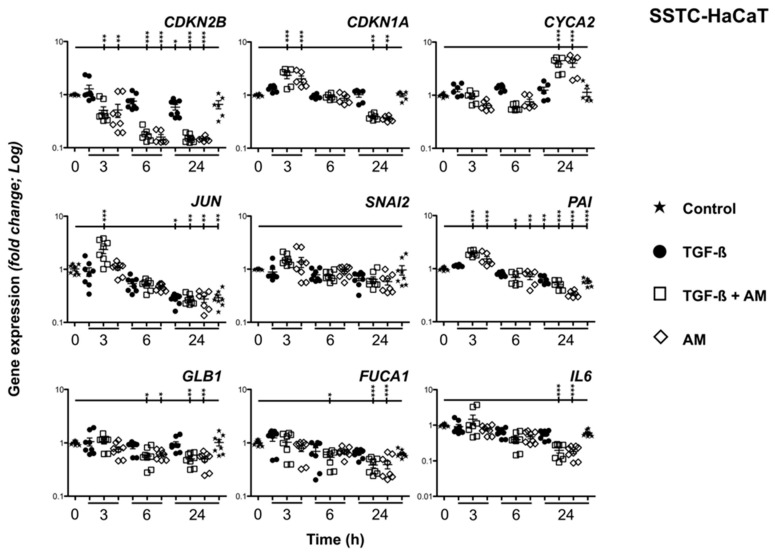
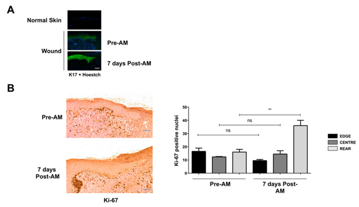
Unsuccessful wound closure in chronic wounds can be linked to altered keratinocyte activation and their inability to re-epithelize. Suggested mechanisms driving this impairment involve unbalanced cytokine signaling. However, the molecular events leading to these aberrant responses are poorly understood. Among cytokines affecting keratinocyte responses, Transforming Growth Factor-β (TFG-β) is thought to have a great impact. In this study, we have used a previously characterized skin epidermal in vitro model, HaCaT cells continuously exposed to TGF-β1, to study the wound recovery capabilities of chronified/senescent keratinocytes. In this setting, chronified keratinocytes show decreased migration and reduced activation in response to injury. Amniotic membrane (AM) has been used successfully to manage unresponsive complicated wounds. In our in vitro setting, AM treatment of chronified keratinocytes re-enabled migration in the early stages of wound healing, also promoting proliferation at later stages. Interestingly, when checking the gene expression of markers known to be altered in TGF-β chronified cells and involved in cell cycle regulation, early migratory responses, senescence, and chronic inflammation, we discovered that AM treatment seemed to reset back to keratinocyte status. The analysis of the evolution of both the levels of keratinocyte activation marker cytokeratin 17 and the spatial-temporal expression pattern of the proliferation marker Ki-67 in human in vivo biopsy samples suggests that responses to AM recorded in TGF-β chronified HaCaT cells would be homologous to those of resident keratinocytes in chronic wounds. All these results provide further evidence that sustained TGF-β might play a key role in wound chronification and postulate the validity of our TGF-β chronified HaCaT in vitro model for the study of chronic wound physiology.

Keywords: TGF-β; amniotic membrane; cell migration; cell models; cell proliferation; chronic wounds; keratinocytes; wound healing.


Conflict of interest statement

The authors declare no conflict of interest. The funders had no role in the design of the study; in the collection, analyses, or interpretation of data; in the writing of the manuscript, or in the decision to publish the results.

Figures

Figure 1
Figure 1
HaCaT cells persistently exposed to TGF-β show decreased migratory capacity. (A) Pictures representative of the migratory performance of SS-HaCaT and SSTC-HaCaT monolayers when challenged with epidermal continuity disruption. (B) Quantification of the migratory performance of SS-HaCaT and SSTC-HaCaT in regard to the initial gap area. Replicates from three independent experiments were analyzed. Shown data represent mean ± SEM. Asterisks denote statistically significant differences (** p < 0.005). The scale bar equals 100 µm.
Figure 2
Figure 2
HaCaT cells persistently exposed to TGF-β showed altered FA dynamics. Distinct spatial and temporal patterns for the structural protein paxillin (red) and the activated modulator factor FAK (green) are shown in both SS-HaCaT and SSTC-HaCaT cells. Nuclei are colored in blue. Representative images of at least three independent experiments are shown. The scale bar equals 50 µm.
Figure 3
Figure 3
Exposure to amniotic membrane empowers migration capabilities and FA dynamics in keratinocytes persistently exposed to TGF-β. (A) Quantification of the migratory performance of SS-HaCaT and SSTC-HaCaT with regard to the initial gap area and the presence of adjuvant treatments. Replicates from three independent experiments were analyzed. Shown data represent the mean ± SEM. Asterisks denote statistically significant differences (* p < 0.05, ** p < 0.005). (B) Spatial and temporal patterns for the structural protein paxillin (red) and the activated modulator factor phosphorylated-FAK (green) in relation to SS-HaCaT and SSTC-HaCaT cell conditions and the exposure to adjuvant treatments. Nuclei are colored blue. Representative images of at least three independent experiments are shown. The scale bar equals 50 µm; ns, non-significant.
Figure 4
Figure 4
Amniotic membrane treatment restricts swelling responses in HaCaT cells persistently exposed to TGF-β. The morphology and size of monolayer cells at the wound edge were studied. Results are shown as fold-change of SSTC-HaCaT cells regarding the same parameters of SS-HaCaT cells and in relation to adjuvant treatments. Replicates from three independent experiments were analyzed. Shown data represent the mean ± SEM. Asterisks denote statistically significant differences (** p < 0.005; **** p < 0.00005); ns, non-significant.
Figure 5
Figure 5
Amniotic membrane promotes cell division at the wound edge of HaCaT-cells persistently exposed to TGF-β. Quantification of the proliferation of wounded in vitro monolayers evidenced through immune labeling. Reference pictures were taken for the indicated times for each condition and treatment. Replicates from three independent experiments were analyzed. Shown data represent the mean ± SEM. Asterisks denote statistically significant differences (* p < 0.05, *** p < 0.0005, **** p < 0.00005); ns, non-significant.
Figure 6
Figure 6
Amniotic membrane treatment reignites the proliferation of keratinocytes despite strong cell-cycle arrest status. (A) Illustrative scheme of the subconfluent HaCaT cells culture setup used. (B) Cell cycle progression was assessed through flow cytometry. Cells were analyzed at the indicated times for each condition and treatment. Replicates from three independent experiments were analyzed and represented. The scale bar equals 100 µm.
Figure 7
Figure 7
Amniotic membrane treatment ameliorates the gene expression pattern of SSTC-HaCaT cells (persistently exposed to TGF-β). Genes related to cell cycle (CDKN2B, CDKN1A, CYCA2), migratory responses (JUN, SNAI2, PAI), and senescent status (GLB1, FUCA1, and IL-6) were studied. Samples were obtained at the indicated times for each condition and treatment. Expression level data are represented as fold change from the initial time. Replicates from three independent experiments were quantified by qPCR. Shown data represent mean ± SEM. Asterisks denote statistically significant differences (* p < 0.05, ** p < 0.005, and *** p < 0.001).
Figure 8
Figure 8
Amniotic membrane treatment reinstates functional activation of HaCaT cells persistently exposed to TGF-β. The ability of keratinocytes to develop proper activation responses upon monolayer disruption was evidenced via keratin-17 (green) immune labeling. Nuclei are colored blue. Representative images taken at the indicated times for each condition and treatment of at least three independent experiments are shown. The scale bar equals 50 µm.
Figure 9
Figure 9
Amniotic membrane effects on chronic wounds correlate well with the observation obtained in vitro on HaCaT persistently exposed to TGF-β. Immune-histological study of human samples obtained from the edge of chronic wounds before and after amniotic membrane treatment. (A) Analysis of the keratinocyte activation using keratin-17 labeling (green). Nuclei are colored blue. (B) Analysis of the keratinocyte proliferation using Ki-67 labeling (brown deposits). Replicates from three independent samples were analyzed. Shown data represent mean ± SEM. Asterisks denote statistically significant differences (** p < 0.005). The scale bar equals 200 µm; ns, non-significant.

References

    1. Singer A.J., Clark R.A. Cutaneous Wound Healing. N. Engl. J. Med. 1999;341:738–746. doi: 10.1056/NEJM199909023411006. - DOI - PubMed
    1. Valluru M., Staton C.A., Reed M.W., Brown N.J. Transforming Growth Factor-Beta and Endoglin Signaling Orchestrate Wound Healing. Front. Physiol. 2011;2:89. doi: 10.3389/fphys.2011.00089. - DOI - PMC - PubMed
    1. Pastar I., Stojadinovic O., Yin N.C., Ramirez H., Nusbaum A.G., Sawaya A., Patel S.B., Khalid L., Isseroff R.R., Tomic-Canic M. Epithelialization in Wound Healing: A Comprehensive Review. Adv. Wound Care. 2014;3:445–464. doi: 10.1089/wound.2013.0473. - DOI - PMC - PubMed
    1. O’Kane S., Ferguson M.W. Transforming Growth Factor Beta S and Wound Healing. Int. J. Biochem. Cell Biol. 1997;29:63–78. doi: 10.1016/S1357-2725(96)00120-3. - DOI - PubMed
    1. Kim B.C., Kim H.T., Park S.H., Cha J.S., Yufit T., Kim S.J., Falanga V. Fibroblasts from Chronic Wounds Show Altered Tgf-Beta-Signaling and Decreased Tgf-Beta Type Ii Receptor Expression. J. Cell Physiol. 2003;195:331–336. doi: 10.1002/jcp.10301. - DOI - PubMed

Substances

LinkOut - more resources