Review
. 2023 Mar 25;24(7):6227.
doi: 10.3390/ijms24076227.

ABC Transporters in Bacterial Nanomachineries

Affiliations
Review

ABC Transporters in Bacterial Nanomachineries

Florestan L Bilsing et al. Int J Mol Sci. .

Abstract

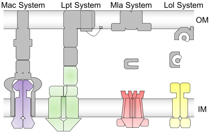
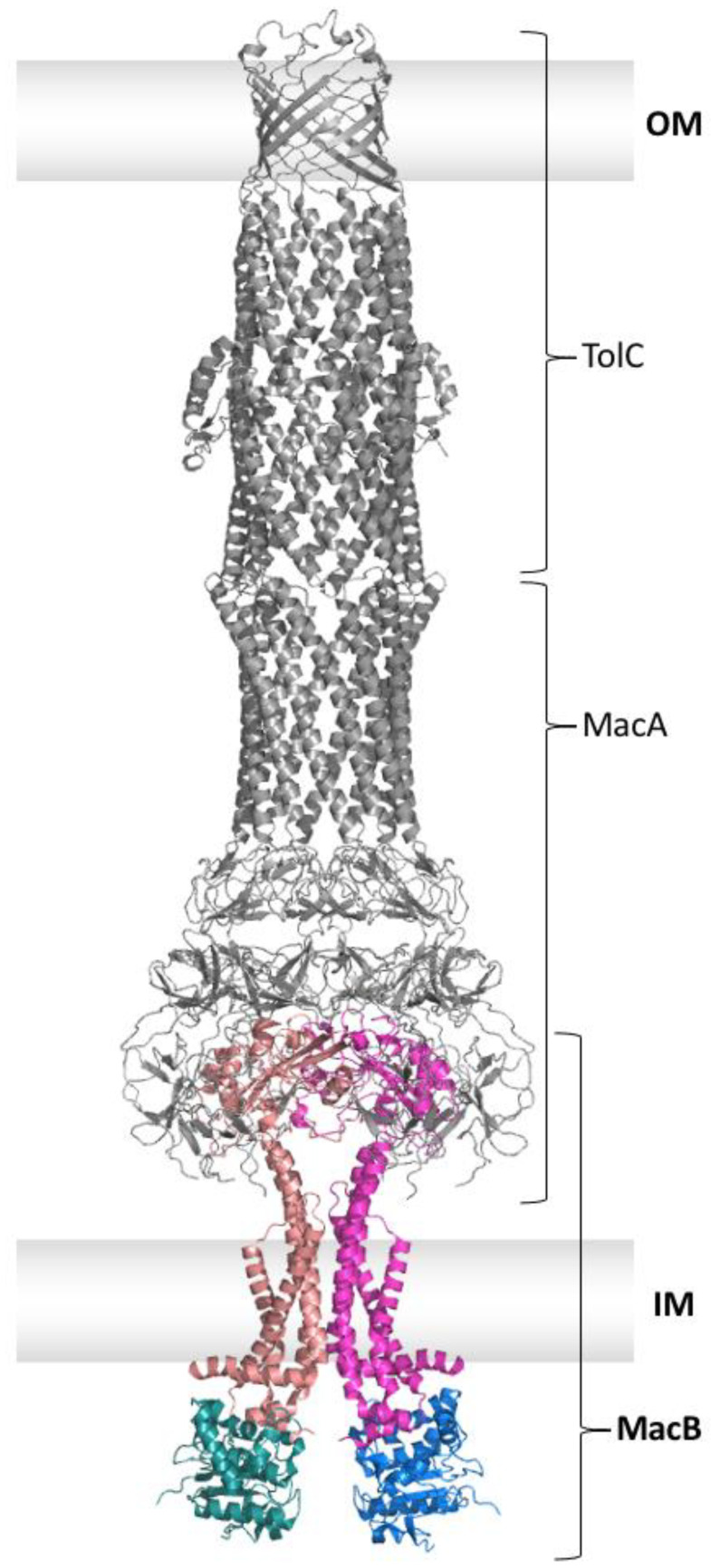
Members of the superfamily of ABC transporters are found in all domains of life. Most of these primary active transporters act as isolated entities and export or import their substrates in an ATP-dependent manner across biological membranes. However, some ABC transporters are also part of larger protein complexes, so-called nanomachineries that catalyze the vectorial transport of their substrates. Here, we will focus on four bacterial examples of such nanomachineries: the Mac system providing drug resistance, the Lpt system catalyzing vectorial LPS transport, the Mla system responsible for phospholipid transport, and the Lol system, which is required for lipoprotein transport to the outer membrane of Gram-negative bacteria. For all four systems, we tried to summarize the existing data and provide a structure-function analysis highlighting the mechanistical aspect of the coupling of ATP hydrolysis to substrate translocation.

Keywords: ABC transporter; Lol system; Lpt system; Mac system; Mla system; nanomachineries.


Conflict of interest statement

The authors declare no conflict of interest.

Figures

Figure 1
Figure 1
Domain organization of prokaryotic ABC exporters. (A): Half-size homodimer. (B): Single domain heterodimer. TMD = transmembrane domain. NBD = nucleotide-binding domain. The TMD and NBD are either fused on one polypeptide chain (A) or exist as separate polypeptides (B).
Figure 2
Figure 2
Schematic of a transport cycle of a type IV ABC exporter. (A): In the inward-facing (IF) conformation, the transporter binds the substrate. (B): During substrate translocation across the membrane, the occluded state is formed with the substrate pocket closed to the inside and outside. (C): The transporter adopts the outward-facing (OF) conformation, and the substrate leaves the transporter to the outside.
Figure 3
Figure 3
Schematic representation of the Mac, Lpt, Mla, and Lol system. For the Mac system, the ABC transporter MacB is shown in purple. The membrane fusion protein MacA and the outer membrane protein TolC are shown in grey. For the Lpt system, the ABC transporter LptB2FGC is shown in green. The periplasmic protein LptA and the translocon in the outer membrane LptDE are shown in grey. For the Mla system, the ABC transporter MlaFEDB is shown in red. The periplasmic protein MlaC and the outer membrane protein MlaA, together with OmpF, are shown in grey. For the Lol system, the ABC transporter LolCDE is shown in yellow. The periplasmic protein LolA and the outer membrane protein LolB are shown in grey. Inner membrane (IM) and outer membrane (OM) are depicted as grey bars. The peptidoglycan layer is omitted for reasons of clarity.
Figure 4
Figure 4
Single particle cryo-EM structure of the assembled MacAB-TolC efflux system (PDB entry 5NIL). Trimeric TolC and hexameric MacA are shown in grey and dimeric MacB in color. The nucleotide-binding domains of MacB are shown in deep teal and marine, and the transmembrane domains, plus the periplasmic part, are shown in salmon and light magenta. The outer membrane (OM) and inner membrane (IM) are displayed as grey boxes. The peptidoglycan layer is omitted for reasons of clarity.
Figure 5
Figure 5
Structures of MacB in a nucleotide-free (PDB entry 5NIL), ATP-bound (PDB entry 5LIL), and ADP-bound (PDB entry 5WS4) state. NBDs are shown in deep teal and marine, and the TMDs with the periplasmic part are shown in salmon and light magenta. (A): Side view of MacB in the different nucleotide-free/-bound states. (B): Top view of the periplasmic part of MacB in the different nucleotide-free/-bound states.
Figure 6
Figure 6
Overview of the Lpt machinery spanning both membranes and the periplasm. Note that the number of LptA oligomers forming the periplasmic bridge is unknown. The figure was created using PyMOL and the PDB entries of LptDE (5IV9, grey in the outer membrane), LptA (2R19, grey in the periplasm), and LptB2FGC (6MJP, orange, salmon, light magenta, deep teal and marine in the inner membrane). The outer membrane (OM) and inner membrane (IM) are displayed as grey boxes. The peptidoglycan layer is omitted for reasons of clarity.
Figure 7
Figure 7
Model and structures of the Lpt systems transport cycle. LptC is shown in orange, while LptF and LptG are shown in light magenta and salmon, respectively. Both LptB protomers are shown in marine and deep teal. LPS is shown in red. Lipids and detergent molecules in the structure are shown as grey sticks, while nucleotides are shown as spheres. (A): Schematic model of LPS extraction by the extractor LptB2FGC. The different states of the transporter are labeled i-v with the PDB entry for the respective structure. i,ii: LPS enters the LptB2FGC cavity from the reader’s side. ii,iii: The LptC TMH leaves the LptFG interface, the cavity tightens and elevates LPS, forming tighter contacts. iii,iv: The LptB2 dimer closes, causing the cavity to collapse and pushes LPS upwards to the β-jellyroll domain of LptF. iv,v: ATP (yellow dots) is hydrolyzed to ADP (grey circles) and Pi, thereby opening LptB2 and the cavity for a new extraction cycle. (B): Crystal structures and single particle cryo-EM structures according to the different states (C): View on the cavity from the periplasmic side (β-jellyroll domains are omitted for clarity). Note that only structures of states ii and iii show LPS. Even though only the structure of state iv shows bound nucleotides, latest data suggest that ATP can bind already during earlier states. Structures of states ii–iv did not resolve the β-jellyroll domain of LptFG. The structure of state ii did not resolve the β-jellyroll domain of LptC, while the structures of states iii–v were lacking LptC completely.
Figure 8
Figure 8
Structural components of the Mla system in a cartoon representation. The homo-trimeric complex of OmpF and MlaA (PDB: 5NUO) situated in the outer membrane is shown in grey color. MlaC (PDB: 6GKI) is shown in the periplasm in grey color. Note: the exact number of MlaC molecules in the periplasm is unknown. The inner membrane complex of MlaFEDB (PDB: 6ZY2) is shown in color: hexameric MlaD, and both MlaB molecules are shown in orange. The two MlaE molecules are shown in salmon and light magenta. The two molecules of MlaF are shown in marine and deep teal. Outer membrane (OM) and inner membrane (IM) are displayed as grey boxes. The peptidoglycan layer is omitted for reasons of clarity.
Figure 9
Figure 9
Structures and model representing the transport cycle of the Mla pathway. MlaC is shown in grey. The hexameric MlaD and both MlaB are shown in orange. MlaE is shown in salmon and light magenta. MlaF is shown in marine and deep teal. Lipid molecules are shown in red, and nucleotides are shown as spheres. The IM is displayed as a grey box. The peptidoglycan layer is omitted for reasons of clarity. (A): Different states of the ABC transporter during the transport cycle: nucleotide-free (PDB: 6ZY2), AMP-PNP-bound (PDB: 6ZY9), ADP-bound(PDB: 6ZY4), and substrate-bound (PDB: 6ZY3). (B): A schematic model of the retrograde transport of the lipid via MlaFEDB. Lipid-loaded MlaC binds to MlaD in the resting state of MlaFEDB. Binding of ATP (yellow dots) prompts the exit of the lipid molecule present in the cavity of MlaE from the last transport cycle. ATP hydrolysis to ADP (grey circles) and Pi prompts dimerization of MlaF and conformational changes in MlaE, which ultimately lead to the extraction of lipid from MlaD-MlaC into the cavity of MlaE. Upon release of hydrolyzed products, the conformation gets back to the resting state.
Figure 10
Figure 10
Overview of the Lol pathway. After insertion of lipoproteins (red, labeled one time with LP) via the Sec or the Tat pathway into the IM, they are diacylated (black) by Lgt in a first modification step. This is followed by cleavage of the N-terminal signal sequence (orange) by Lsp. Subsequently, Lnt acylates the newly N-terminally located cysteine, which makes the now mature lipoprotein ready for transport via the Lol machinery. This starts with the extraction of the lipoprotein from the cytoplasmic membrane via the ABC transporter LolCDE (salmon, light magenta, deep teal, and marine), which leads to the delivery of the lipoprotein to the periplasmic chaperone LolA (grey). LolA shuffles the lipoprotein to the last checkpoint LolB (grey), which finally inserts the lipoprotein into the outer membrane.
Figure 11
Figure 11
Structures of LolCDE in the Apo (PDB: 7V8M), lipoprotein-bound (PDB: 7V8L), and AMP-PNP-bound (PDB: 7V8I) form. LolC is shown in salmon, LolE is shown in light magenta, one LolD monomer is shown in deep teal, and the other LolD monomer is shown in marine. The lipoprotein is shown in red spheres. (A): Side-view of LolCDE. In the Apo state, LolCDE exhibits a V-shaped cavity that the lipoprotein enters from the membrane. Upon ATP binding (AMP-PNP-bound state), the central cavity is closed, and the substrate is shuffled out of LolCDE to LolA. (B): Cross-sectional view of the TMDs of LolCDE in the different substrate- or nucleotide-bound states. Movement of TMH2 of LolE upon ATP-binding into the central cavity extrudes the substrate out of the transporter.

Similar articles

Cited by

References

    1. Higgins C.F. ABC transporters: Physiology, structure and mechanism--an overview. Res. Microbiol. 2001;152:205–210. doi: 10.1016/S0923-2508(01)01193-7. - DOI - PubMed
    1. Davidson A.L., Dassa E., Orelle C., Chen J. Structure, function, and evolution of bacterial ATP-binding cassette systems. Microbiol. Mol. Biol. Rev. 2008;72:317–364. doi: 10.1128/MMBR.00031-07. - DOI - PMC - PubMed
    1. Biemans-Oldehinkel E., Doeven M.K., Poolman B. ABC transporter architecture and regulatory roles of accessory domains. FEBS Lett. 2006;580:1023–1035. doi: 10.1016/j.febslet.2005.11.079. - DOI - PubMed
    1. Thomas C., Tampe R. Structural and Mechanistic Principles of ABC Transporters. Annu. Rev. Biochem. 2020;89:605–636. doi: 10.1146/annurev-biochem-011520-105201. - DOI - PubMed
    1. Oldham M.L., Davidson A.L., Chen J. Structural insights into ABC transporter mechanism. Curr. Opin. Struct. Biol. 2008;18:726–733. doi: 10.1016/j.sbi.2008.09.007. - DOI - PMC - PubMed

MeSH terms

Substances

LinkOut - more resources