

Crystal Structure of Pyrrolysyl-tRNA Synthetase from a Methanogenic Archaeon ISO4-G1 and Its Structure-Based Engineering for Highly-Productive Cell-Free Genetic Code Expansion with Non-Canonical Amino Acids
- PMID: 37047230
- PMCID: PMC10094482
- DOI: 10.3390/ijms24076256
Crystal Structure of Pyrrolysyl-tRNA Synthetase from a Methanogenic Archaeon ISO4-G1 and Its Structure-Based Engineering for Highly-Productive Cell-Free Genetic Code Expansion with Non-Canonical Amino Acids
Abstract
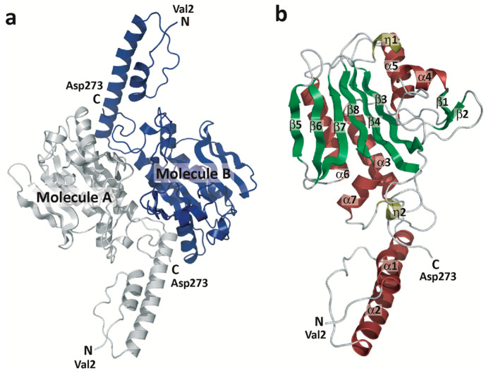
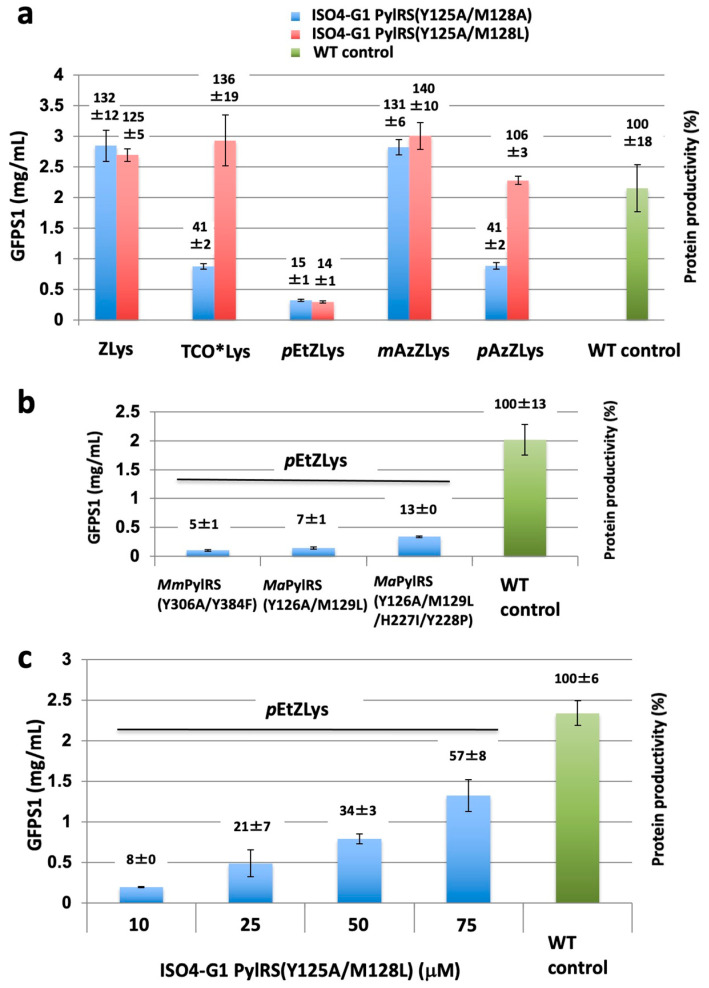
Pairs of pyrrolysyl-tRNA synthetase (PylRS) and tRNAPyl from Methanosarcina mazei and Methanosarcina barkeri are widely used for site-specific incorporations of non-canonical amino acids into proteins (genetic code expansion). Previously, we achieved full productivity of cell-free protein synthesis for bulky non-canonical amino acids, including Nε-((((E)-cyclooct-2-en-1-yl)oxy)carbonyl)-L-lysine (TCO*Lys), by using Methanomethylophilus alvus PylRS with structure-based mutations in and around the amino acid binding pocket (first-layer and second-layer mutations, respectively). Recently, the PylRS·tRNAPyl pair from a methanogenic archaeon ISO4-G1 was used for genetic code expansion. In the present study, we determined the crystal structure of the methanogenic archaeon ISO4-G1 PylRS (ISO4-G1 PylRS) and compared it with those of structure-known PylRSs. Based on the ISO4-G1 PylRS structure, we attempted the site-specific incorporation of Nε-(p-ethynylbenzyloxycarbonyl)-L-lysine (pEtZLys) into proteins, but it was much less efficient than that of TCO*Lys with M. alvus PylRS mutants. Thus, the first-layer mutations (Y125A and M128L) of ISO4-G1 PylRS, with no additional second-layer mutations, increased the protein productivity with pEtZLys up to 57 ± 8% of that with TCO*Lys at high enzyme concentrations in the cell-free protein synthesis.
Keywords: cell-free protein synthesis; crystal structure; genetic code expansion; non-canonical amino acids; tRNA.
Conflict of interest statement
T.Y., E.S., K.S., and S.Y. are co-inventors on the patent [WO2020/045656 A1] related to this work. S.Y. is a founder and shareholder of LiberoThera Co., Ltd.
Figures


Similar articles
-
Fully Productive Cell-Free Genetic Code Expansion by Structure-Based Engineering of Methanomethylophilus alvus Pyrrolysyl-tRNA Synthetase.ACS Synth Biol. 2020 Apr 17;9(4):718-732. doi: 10.1021/acssynbio.9b00288. Epub 2020 Mar 17. ACS Synth Biol. 2020. PMID: 32182048
-
Generating Efficient Methanomethylophilus alvus Pyrrolysyl-tRNA Synthetases for Structurally Diverse Non-Canonical Amino Acids.ACS Chem Biol. 2022 Dec 16;17(12):3458-3469. doi: 10.1021/acschembio.2c00639. Epub 2022 Nov 16. ACS Chem Biol. 2022. PMID: 36383641 Free PMC article.
-
Structural Basis for Genetic-Code Expansion with Bulky Lysine Derivatives by an Engineered Pyrrolysyl-tRNA Synthetase.Cell Chem Biol. 2019 Jul 18;26(7):936-949.e13. doi: 10.1016/j.chembiol.2019.03.008. Epub 2019 Apr 25. Cell Chem Biol. 2019. PMID: 31031143
-
Update of the Pyrrolysyl-tRNA Synthetase/tRNAPyl Pair and Derivatives for Genetic Code Expansion.J Bacteriol. 2023 Feb 22;205(2):e0038522. doi: 10.1128/jb.00385-22. Epub 2023 Jan 25. J Bacteriol. 2023. PMID: 36695595 Free PMC article. Review.
-
tRNAPyl: Structure, function, and applications.RNA Biol. 2018;15(4-5):441-452. doi: 10.1080/15476286.2017.1356561. Epub 2017 Sep 13. RNA Biol. 2018. PMID: 28837402 Free PMC article. Review.
Cited by
-
Engineering Pyrrolysine Systems for Genetic Code Expansion and Reprogramming.Chem Rev. 2024 Oct 9;124(19):11008-11062. doi: 10.1021/acs.chemrev.4c00243. Epub 2024 Sep 5. Chem Rev. 2024. PMID: 39235427 Free PMC article. Review.
-
An efficient pyrrolysyl-tRNA synthetase for economical production of MeHis-containing enzymes.Faraday Discuss. 2024 Sep 11;252(0):295-305. doi: 10.1039/d4fd00019f. Faraday Discuss. 2024. PMID: 38847587 Free PMC article.
-
Beyond In Vivo, Pharmaceutical Molecule Production in Cell-Free Systems and the Use of Noncanonical Amino Acids Therein.Chem Rev. 2025 Feb 12;125(3):1303-1331. doi: 10.1021/acs.chemrev.4c00126. Epub 2025 Jan 22. Chem Rev. 2025. PMID: 39841856 Free PMC article. Review.
-
Pyrrolysine Aminoacyl-tRNA Synthetase as a Tool for Expanding the Genetic Code.Int J Mol Sci. 2025 Jan 10;26(2):539. doi: 10.3390/ijms26020539. Int J Mol Sci. 2025. PMID: 39859254 Free PMC article. Review.
References
MeSH terms
Substances
Grants and funding
- The Platform Project for Supporting Drug Discovery and Life Science Research (Basis for Sup-porting Innovative Drug Discovery and Life Science Research (BINDS)) under Grant Number JP17am0101081/Japan Agency for Medical Research and Development
- Leading Advanced Projects for Medical Innovation (LEAP) under Grant Number JP19gm0010001/Japan Agency for Medical Research and Development
- N/A/Takeda Science Foundation
- 16K05859/Ministry of Education, Culture, Sports, Science and Technology
- 24550203/Ministry of Education, Culture, Sports, Science and Technology
LinkOut - more resources
Full Text Sources
Medical
Research Materials
