Review
. 2023 Mar 27;24(7):6301.
doi: 10.3390/ijms24076301.

Chitinase Signature in the Plasticity of Neurodegenerative Diseases

Affiliations
Review

Chitinase Signature in the Plasticity of Neurodegenerative Diseases

Cristina Russo et al. Int J Mol Sci. .

Abstract

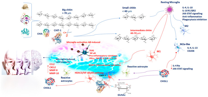
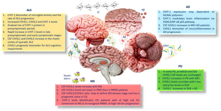
Several reports have pointed out that Chitinases are expressed and secreted by various cell types of central nervous system (CNS), including activated microglia and astrocytes. These cells play a key role in neuroinflammation and in the pathogenesis of many neurodegenerative disorders. Increased levels of Chitinases, in particular Chitotriosidase (CHIT-1) and chitinase-3-like protein 1 (CHI3L1), have been found increased in several neurodegenerative disorders. Although having important biological roles in inflammation, to date, the molecular mechanisms of Chitinase involvement in the pathogenesis of neurodegenerative disorders is not well-elucidated. Several studies showed that some Chitinases could be assumed as markers for diagnosis, prognosis, activity, and severity of a disease and therefore can be helpful in the choice of treatment. However, some studies showed controversial results. This review will discuss the potential of Chitinases in the pathogenesis of some neurodegenerative disorders, such as Alzheimer's disease, Parkinson's disease, amyotrophic lateral sclerosis, and multiple sclerosis, to understand their role as distinctive biomarkers of neuronal cell activity during neuroinflammatory processes. Knowledge of the role of Chitinases in neuronal cell activation could allow for the development of new methodologies for downregulating neuroinflammation and consequently for diminishing negative neurological disease outcomes.

Keywords: Alzheimer’s disease; Chitotriosidase; Parkinson’s disease; amyotrophic lateral sclerosis; chitinase-3-like 1; glycohydrolase family 18; multiple sclerosis; neuroinflammation.


Conflict of interest statement

The authors declare to not have any potential conflict of interest.

Figures

Figure 1
Figure 1
Chitin has both pro-inflammatory and anti-inflammatory effects on the basis of its molecular size: BC (>70 µm) is inert to the inflammatory response; SC (<40 µm) exerts an anti-inflammatory response via IL-10 production. IL-10, binding with its own receptors IL-10R1 and IL-10R2, triggers the JAK/STAT signaling pathway, resulting in pro-inflammatory cytokine and phagocytosis inhibition; IC (40–70 µm) induces pro-inflammatory responses via TLR2- and NF-κB-dependent pathways for IFN-γ, TNF-α, IL1, IL2, and IL12 production. Microglial cells engulfing chitin debris, GlcNAc polymers, and Aβ induce NLRP3 inflammasome activation and neurotoxicity. CHIT-1 and CHIA are the most important active enzymes able to degrade chitin polymers. CHIT-1 modulates neuroinflammation via the HDAC3/NF-κB p65 pathway contributing to AD progression. Microglia polarize in M1 and M2, displaying pro-inflammatory and anti-inflammatory phenotypes, respectively. The M1-polarized phenotype promotes neuronal damage, whereas the M2-polarized phenotype is immunosuppressive and neuroprotective. M2a-like microglia induce CHI3L1, IL13, and CD206. CHI3L1 modulates IL-4Rα expression and STAT6 phosphorylation, affecting M2 polarization. CHI3L1 is expressed in microglia, infiltrating macrophages and astrocytes. The expression of CHI3L1 is induced by pro-inflammatory cytokines, including IL-6, IFN-γ, IL-1β, and TNF-α. Its regulation by IL-6 and TNF-α needs NF-κB activation. CHI3L1 is induced by inflammatory proteins, such as MMP-10, CX3CL1, and 4E-BP1, and therefore, its activity facilitates AD neuroinflammation. Blue arrows represent induction; red arrows represent inhibition. Abbreviation= BC, IC, SC: big, intermediate, small, chitin; IL: interleukin; TLR2: Toll–like receptor 2; NF-κB: nuclear factor kappa-light-chain-enhancer of activated B cells; IFN-γ: interferon-gamma; TNF-α: tumor necrosis factor-α; GlcNAc: β-1,4-linked N-acetylglucosamine; Aβ: beta-amyloid protein; NLRP3: NLR Family Pyrin Domain Containing 3; CHIT-1: Chitotriosidase; CHIA: chitinase acid; AD: Alzheimer’s disease; CHI3L1: chitinase-3-like protein 1; MMP: marrow matrix metalloproteinase; CXCL: Chemokine family; 4E-BP1: eukaryotic translation initiation factor 4E-binding protein 1.
Figure 2
Figure 2
Schematic representation of the chronological profiles of Chitinases in AD, PD, ALS, and progression. Abbreviation = AD: Alzheimer’s disease; PD: Parkinson’s disease; ALS: amyotrophic lateral sclerosis; MS: multiple sclerosis; CSF: cerebrospinal fluid; CHI3L1: chitinase-3-like protein 1; CHI3L2: chitinase-3-like 2; MCI: mild cognitive impairment; CIS: clinically isolated syndrome; DLB: dementia with Lewy bodies; prodDLB: prodromal dementia with Lewy bodies.

Similar articles

Cited by

References

    1. Gitler A.D., Dhillon P., Shorter J. Neurodegenerative disease: Models, mechanisms, and a new hope. Dis. Model Mech. 2017;10:499–502. doi: 10.1242/dmm.030205. - DOI - PMC - PubMed
    1. Dugger B.N., Dickson D.W. Pathology of Neurodegenerative Diseases. Cold Spring Harb. Perspect. Biol. 2017;9:028035. doi: 10.1101/cshperspect.a028035. - DOI - PMC - PubMed
    1. Dharshini S.A.P., Jemimah S., Taguchi Y.H., Gromiha M.M. Exploring Common Therapeutic Targets for Neurodegenerative Disorders Using Transcriptome Study. Front. Genet. 2021;12:639160. doi: 10.3389/fgene.2021.639160. - DOI - PMC - PubMed
    1. Kovacs G.G. Pathology of Neurodegenerative Diseases. J. Clin. Pathol. 2019;72:725–735. doi: 10.1136/jclinpath-2019-205952. - DOI - PubMed
    1. Malaguarnera L. Chitotriosidase: The yin and yang. Cell. Mol. Life Sci. 2006;63:3018–3029. doi: 10.1007/s00018-006-6269-2. - DOI - PMC - PubMed