. 2023 Mar 28;24(7):6322.
doi: 10.3390/ijms24076322.

IL-10 Promotes CXCL13 Expression in Macrophages Following Foot-and-Mouth Disease Virus Infection

Affiliations

IL-10 Promotes CXCL13 Expression in Macrophages Following Foot-and-Mouth Disease Virus Infection

Zijing Guo et al. Int J Mol Sci. .

Abstract

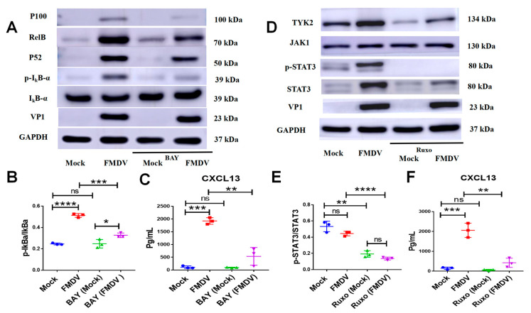
Foot-and-mouth disease (FMD) is one of the most contagious livestock diseases in the world, posing a constant global threat to the animal trade and national economies. The chemokine C-X-C motif chemokine ligand 13 (CXCL13), a biomarker for predicting disease progression in some diseases, was recently found to be increased in sera from mice infected with FMD virus (FMDV) and to be associated with the progression and severity of the disease. However, it has not yet been determined which cells are involved in producing CXCL13 and the signaling pathways controlling CXCL13 expression in these cells. In this study, the expression of CXCL13 was found in macrophages and T cells from mice infected with FMDV, and CXCL13 was produced in bone-marrow-derived macrophages (BMDMs) by activating the nuclear factor-kappaB (NF-κB) and JAK/STAT pathways following FMDV infection. Interestingly, CXCL13 concentration was decreased in sera from interleukin-10 knock out (IL-10-/-) mice or mice blocked IL-10/IL-10R signaling in vivo after FMDV infection. Furthermore, CXCL13 was also decreased in IL-10-/- BMDMs and BMDMs treated with anti-IL-10R antibody following FMDV infection in vitro. Lastly, it was demonstrated that IL-10 regulated CXCL13 expression via JAK/STAT rather than the NF-κB pathway. In conclusion, the study demonstrated for the first time that macrophages and T cells were the cellular sources of CXCL13 in mice infected with FMDV; CXCL13 was produced in BMDMs via NF-κB and JAK/STAT pathways; and IL-10 promoted CXCL13 expression in BMDMs via the JAK/STAT pathway.

Keywords: C-X-C motif chemokine ligand 13; foot-and-mouth disease virus; interleukin-10; macrophage; signaling pathways.


Conflict of interest statement

The authors declare that the study was conducted in the absence of any commercial or financial relationships that could be construed as a potential conflict of interest.

Figures

Figure 1
Figure 1
Macrophages and T cells were the cellular sources of CXCL13 in FMDV-infected mice. (A) The mRNA expression of CXCL13 in various tissues from mock mice and infected mice at 12, 24, 36, 48, 60, and 72 hpi. (B) The mRNA expression of CXCL13 in various cell subsets sorted from mock mice and infected mice. CXCL13, F4/80 (C) or CD3 (D), and nucleus were co-stained in the spleen from mock mice. CXCL13, F4/80 (E) or CD3 (F), and nucleus were co-stained in the spleen from infected mice. CXCL13, F4/80, CD3, and nucleus are shown in green, red, red and blue, respectively. At least 10 fields of immunofluorescence were observed. Significance was detected using unpaired t test. *, p ≤ 0.05; **, p ≤ 0.01. While ns indicates a non-statistically significant difference (p > 0.05).
Figure 2
Figure 2
CXCL13 was produced in BMDMs after FMDV infection. (A) The mRNA expressions and the protein levels of CXCL13 in uninfected BMDMs (Mock) and FMDV-infected BMDMs at 24 hpi were quantified by RT-qPCR and ELISA, respectively. (B) CXCL13 (green), F4/80 (red), and nucleus (blue) were co-stained in Mock and FMDV-infected BMDMs at 24 hpi. (C) The FMDV RdRp sequence and FMDV VP1 protein were detected in Mock and FMDV-infected BMDMs at 24 hpi with RT-qPCR and Western blotting, respectively. Upper panels, original magnification ×100 (scale bar = 25 μm). The white box indicated the regions at higher magnifications (scale bar = 7.5 μm). At least 10 fields of immunofluorescence were observed. Significance was detected using unpaired t test. *, p ≤ 0.05; ***, p ≤ 0.001.
Figure 3
Figure 3
IL-10 mediated CXCL13 expression in FMDV-infected mice. (A) CXCL13 concentrations in sera from mock mice, infected mice with anti-IL-10R antibody, and infected mice with isotype antibody were measured by ELISA. (B) CXCL13 concentrations in sera from mock mice, wild-type mice infected with FMDV, IL-10-/- mice, and IL-10-/- mice infected with FMDV were measured by ELISA. The mRNA expressions of CXCL13 were detected in the heart (C) and spleen (D) of mock mice, infected-mice with anti-IL-10R antibody, and infected-mice with isotype antibody at 48 hpi. The mRNA expressions of CXCL13 were detected in the heart (E) and spleen (F) of mock mice, wild-type mice infected with FMDV, IL-10-/- mice, and IL-10-/- mice infected with FMDV at 48 hpi. Significance was detected using unpaired t test. **, p ≤ 0.01; ***, p ≤ 0.001; ****, p ≤ 0.0001.
Figure 4
Figure 4
IL-10 controlled CXCL13 expression in BMDMs infected with FMDV. The mRNA (A) and protein (B) expression of IL-10 in BMDMs infected with FMDV at 24 hpi were measured by RT-qPCR and ELISA, respectively. The CXCL13 concentrations of the supernatant from BMDMs, BMDMs infected with FMDV, BMDMs treated by anti-IL-10R antibody and infected with FMDV, and BMDMs stimulated with rIL-10 at 20 ng/mL (C) and from BMDMs, BMDMs infected with FMDV, IL-10-/- BMDMs, and IL-10-/- BMDMs infected with FMDV at 24 hpi (D) were measured by ELISA. Significance was detected using unpaired t test. *, p ≤ 0.05; **, p ≤ 0.01; ***, p ≤ 0.001. While ns indicates a non-statistically significant difference (p > 0.05).
Figure 5
Figure 5
FMDV induces CXCL13 production in BMDMs by activating NF-κB and JAK/STAT signaling pathways. (A) The expressions of VP1, IκBa, p-IκBa, P100, P52, RelB and glyceraldehyde-3-phosphate dehydrogenase (GAPDH) in cell lysates of BMDMs, BMDMs infected with FMDV, BMDMs treated by BAY at the recommended concentration of 10 μM, and BMDMs treated by BAY and FMDV were analyzed in by Western blotting. (B) The gray value ratio of p-IκBa/IκBa in BMDMs under different conditions of BAY treatment and FMDV infection. (C) The CXCL13 concentrations of the supernatant from BMDMs under different conditions of BAY treatment and FMDV infection were measured by ELISA. (D) The expressions of VP1, STAT3, p-STAT3, JAK1, TYK2 and GAPDH in cell lysates of BMDMs, BMDMs infected with FMDV, BMDMs treated by ruxolitinib at the recommended concentration of 10 μM, and BMDMs treated by ruxolitinib and FMDV were analyzed by Western blotting. (E) The analysis of the gray value ratio of p-STAT3/STAT3 in BMDMs under different conditions of ruxolitinib treatment and FMDV infection. (F) The CXCL13 concentrations of the supernatant from BMDMs under different conditions of ruxolitinib treatment and FMDV infection were measured by ELISA. Significance was detected using unpaired t test. *, p ≤ 0.05; **, p ≤ 0.01; ***, p ≤ 0.001; ****, p ≤ 0.0001. While ns indicates a non-statistically significant difference (p > 0.05).
Figure 6
Figure 6
IL-10 induces CXCL13 expression in BMDMs by activating JAK/STAT signaling pathway. (A) The CXCL13 concentration in BMDMs followed the rIL-10 induction and treated with BAY or ruxolitinib at the same time. The expressions of VP1, P52, p-P65, RelB, P100, STAT3, p-STAT3, TYK2 and GAPDH in cell lysates of BMDMs, BMDMs infected with FMDV, BMDMs treated by anti-IL-10R antibody and infected with FMDV, and BMDMs stimulated with rIL-10 at 20 ng/mL (B) and in cell lysates of BMDMs, BMDMs infected with FMDV, IL-10-/-BMDMs, and IL-10-/- BMDMs infected with FMDV (C) were analyzed by Western blotting. The gray value ratio of p-STAT3/STAT3 in BMDMs, BMDMs infected with FMDV, BMDMs treated by anti-IL-10R antibody and infected with FMDV, and BMDMs stimulated with rIL-10 at 20 ng/mL (D) and in BMDMs, BMDM infected with FMDV, IL-10-/- BMDMs, and IL-10-/- BMDMs infected with FMDV at 24 hpi (E). Significance was detected using unpaired t test. ***, p ≤ 0.001. While ns indicates a non-statistically significant difference (p > 0.05).

References

    1. Kazanietz M.G., Durando M., Cooke M. CXCL13 and Its Receptor CXCR5 in Cancer: Inflammation, Immune Response, and Beyond. Front. Endocrinol. 2019;10:471. doi: 10.3389/fendo.2019.00471. - DOI - PMC - PubMed
    1. Bekele Feyissa Y., Chiodi F., Sui Y., Berzofsky J.A. The Role of CXCL13 in Antibody Responses to HIV-1 Infection and Vaccination. Front. Immunol. 2021;12:638872. doi: 10.3389/fimmu.2021.638872. - DOI - PMC - PubMed
    1. Tsai C.H., Chen C.J., Gong C.L., Liu S.C., Chen P.C., Huang C.C., Hu S.L., Wang S.W., Tang C.H. CXCL13/CXCR5 axis facilitates endothelial progenitor cell homing and angiogenesis during rheumatoid arthritis progression. Cell Death Dis. 2021;12:846. doi: 10.1038/s41419-021-04136-2. - DOI - PMC - PubMed
    1. Litsiou E., Semitekolou M., Galani I.E., Morianos I., Tsoutsa A., Kara P., Rontogianni D., Bellenis I., Konstantinou M., Potaris K., et al. CXCL13 production in B cells via Toll-like receptor/lymphotoxin receptor signaling is involved in lymphoid neogenesis in chronic obstructive pulmonary disease. Am. J. Respir. Crit. Care Med. 2013;187:1194–1202. doi: 10.1164/rccm.201208-1543OC. - DOI - PubMed
    1. Wang B., Wang M., Ao D., Wei X. CXCL13-CXCR5 axis: Regulation in inflammatory diseases and cancer. Biochim. Biophys. Acta. Rev. Cancer. 2022;1877:188799. doi: 10.1016/j.bbcan.2022.188799. - DOI - PubMed

LinkOut - more resources