Review
. 2023 Mar 28;24(7):6334.
doi: 10.3390/ijms24076334.

Specialized Ribosomes in Health and Disease

Affiliations
Review

Specialized Ribosomes in Health and Disease

Sarah C Miller et al. Int J Mol Sci. .

Abstract

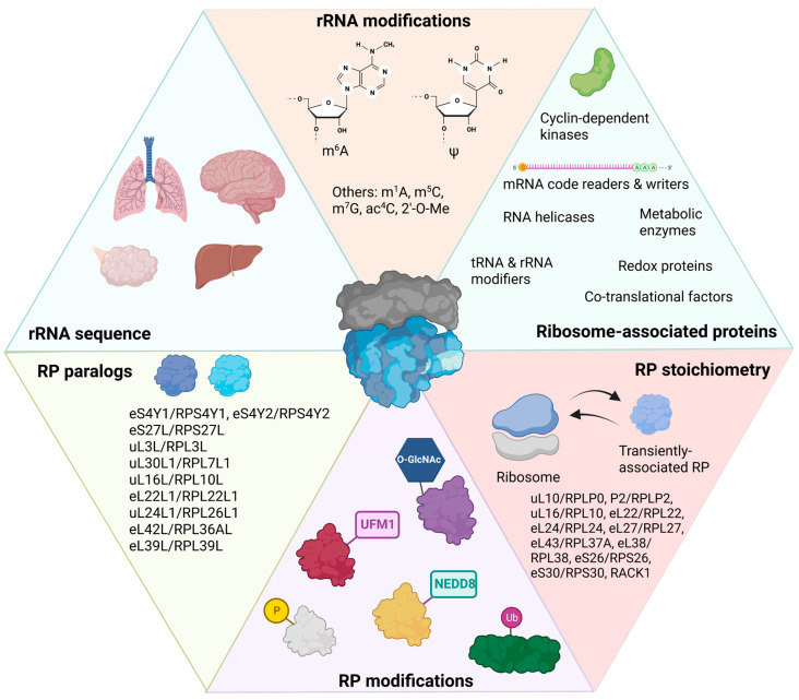
Ribosomal heterogeneity exists within cells and between different cell types, at specific developmental stages, and occurs in response to environmental stimuli. Mounting evidence supports the existence of specialized ribosomes, or specific changes to the ribosome that regulate the translation of a specific group of transcripts. These alterations have been shown to affect the affinity of ribosomes for certain mRNAs or change the cotranslational folding of nascent polypeptides at the exit tunnel. The identification of specialized ribosomes requires evidence of the incorporation of different ribosomal proteins or of modifications to rRNA and/or protein that lead(s) to physiologically relevant changes in translation. In this review, we summarize ribosomal heterogeneity and specialization in mammals and discuss their relevance to several human diseases.

Keywords: human disease; protein synthesis; ribosomal RNA; ribosomal protein; ribosome heterogeneity; ribosome specialization; translation; translational control.


Conflict of interest statement

The authors declare no conflict of interest.

Figures

Figure 1
Figure 1
Ribosomal heterogeneity in mammals. Ribosomal RNA (rRNA) contains tissue-specific sequences (blue panel). There are several rRNA modifications, including N6-methyladenosine (m6A), N1-methyladenosine (m1A), pseudouridine (Ψ), 5-methylcyosine (m5C), N7-methylguanosine (m7G), N4-acetylation of cytosine (ac4C), and 2′-O-methylation (2′-O-Me) (orange panel). Multiple factors associate with ribosomes, altering their translational capacities (green panel). Ribosomal protein (RP) abundance can affect ribosome stoichiometry, and some RPs are known to be incorporated into ribosomes outside of canonical ribosome biogenesis in neurons (red panel). RPs can also contain several post-translational modifications: “P” is phosphorylation; “Ub” is ubiquitin protein; “UFM1” is ubiquitin-fold modifier 1 protein; “NEDD8” is neural precursor cell-expressed, developmentally down-regulated 8 protein, a Ub-like protein; and “O-GlcNAc” is O-linked β-N-acetylglucosamine (purple panel). Nine canonical RPs also have paralogous proteins, some of which are tissue specific (yellow panel). This figure was created with BioRender.com.
Figure 2
Figure 2
Locations of ribosomal proteins that potentially contribute to specialization. (A) The mRNA entry tunnel RPs are indicated: eS30/RPS30, eS12/RPS12, uS3/RPS3, and uS7/RPS5. The E-site of PDB 6QZP is indicated by a yellow tRNA for orientation. Both 40S (light gray) and 60S (cyan) subunits are indicated. The ribosome has the position of −y and is indicated by the E-site tRNA on the right-hand side of the image. (B) The mRNA exit tunnel RPs are indicated: uS11/RPS14, eS28/RPS28, eS26/RPS26, and eS1/RPS3A. The ribosome has the position of +y and is indicated by the E-site tRNA on the left-hand side of the image. Panel (A) is 180 degrees from (B). (C) The nascent polypeptide exit tunnel (NPET) RPs are indicated in different colors: eL19/RPL19, uL23/RPL23A, uL22/RPL22, uL29/RPL35, uL24/RPL26, uL4/RPL4, and eL39/RPL39. Both the +y and −y ribosome directions are indicated by the 180° turn, with an additional 90° turn (total 270° degrees) to better show eL39/RPL39. (D) The locations of RPs in tRNA binding pockets are indicated by different colors: eL27/RPL27, uS12/RPS23, eS25/RPS25, uL5/RPL11, and uL16/RPL10. The E-site is indicated as before for orientation, as well as the 40S and 60S proteins. This figure was created using PyMOL.
Figure 3
Figure 3
Locations of ribosomal proteins that are implicated in ribosome specialization. (A) 40S ribosomal proteins are indicated by different colors: eS26/RPS26, eS27/RPS27, eS6/RPS6, eS24/RPS24, and eS19/RPS19. The E-site of PDB 6QZP is indicated by a yellow tRNA for orientation. Both the +y and −y ribosome directions are indicated by the 180° turn. Both 40S (light gray) and 60S (cyan) are indicated. (B) 60S ribosomal proteins are indicated by different colors: uL1/RPL10A, eL42/RPL36A, eL18/RPL18, eL15/RPL15, eL28/RPL28, uL3/RPL3, eL38/RPL38, eL22/RPL22, uL13/RPL13A, uL30/RPL7, eL20/RPL18A, and eL40/RPL40. The E-site and ribosome directions are indicated as above. (C) X-linked RPs are represented by different colors: eS4X/RPS4X, eL39/RPL39, eL42/RPL36A, and uL16L/RPL10L. 28S has been hidden to show the interior proteins uL16L/RPL10L and eL42/RPL36A. The E-site, directionality, and ribosome subunits are indicated as above. This figure was created using PyMOL.
Figure 4
Figure 4
Specialized ribosomal proteins are expressed in male germ cells in mammals. (A) Locations of RPs and RP-associated protein genes on the human X chromosome. (B) Timeline of mouse spermatogenesis with periods of male sex chromosome inactivation (MSCI) indicated. Times are in days. Colored bars along gene names indicate approximate periods of expression of either the X-linked genes or their homologs in human and mouse male germ cells.

Similar articles

Cited by

References

    1. Geiger T., Wehner A., Schaab C., Cox J., Mann M. Comparative Proteomic Analysis of Eleven Common Cell Lines Reveals Ubiquitous but Varying Expression of Most Proteins. Mol. Cell. Proteom. 2012;11:11. doi: 10.1074/mcp.M111.014050. - DOI - PMC - PubMed
    1. Genuth N.R., Barna M. The Discovery of Ribosome Heterogeneity and Its Implications for Gene Regulation and Organismal Life. Mol. Cell. 2018;71:364–374. doi: 10.1016/j.molcel.2018.07.018. - DOI - PMC - PubMed
    1. Kampen K.R., Sulima S.O., Vereecke S., De Keersmaecker K. Hallmarks of ribosomopathies. Nucleic Acids Res. 2020;48:1013–1028. doi: 10.1093/nar/gkz637. - DOI - PMC - PubMed
    1. Guimaraes J.C., Zavolan M. Patterns of ribosomal protein expression specify normal and malignant human cells. Genome Biol. 2016;17:236. doi: 10.1186/s13059-016-1104-z. - DOI - PMC - PubMed
    1. Panda A., Yadav A., Yeerna H., Singh A., Biehl M., Lux M., Schulz A., Klecha T., Doniach S., Khiabanian H., et al. Tissue- and development-stage-specific mRNA and heterogeneous CNV signatures of human ribosomal proteins in normal and cancer samples. Nucleic Acids Res. 2020;48:7079–7098. doi: 10.1093/nar/gkaa485. - DOI - PMC - PubMed

LinkOut - more resources