

Efficacy and Microbiota Modulation Induced by LimpiAL 2.5%, a New Medical Device for the Inverse Psoriasis Treatment
- PMID: 37047310
- PMCID: PMC10094358
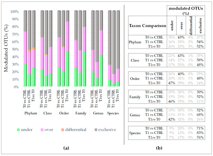
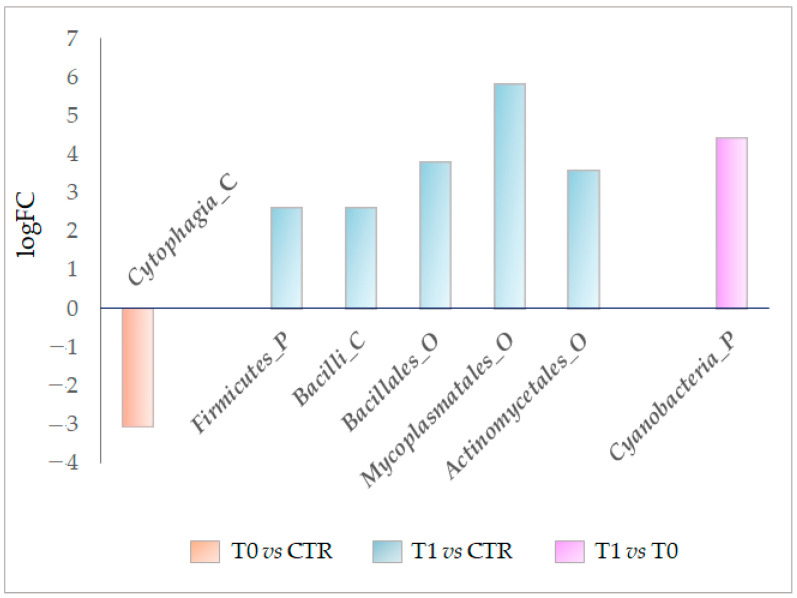
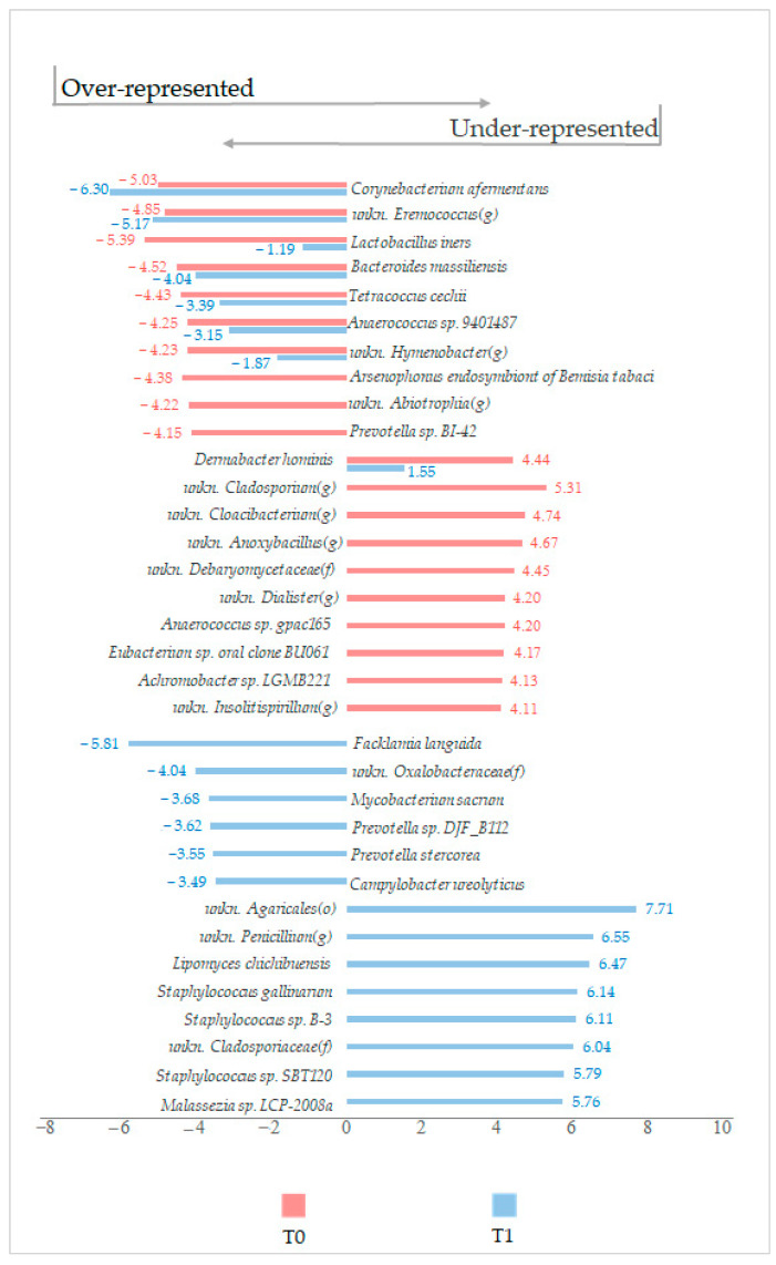
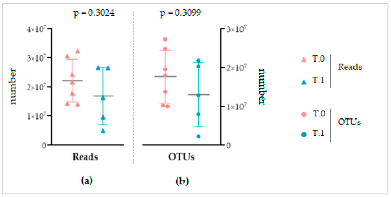
- DOI: 10.3390/ijms24076339
Efficacy and Microbiota Modulation Induced by LimpiAL 2.5%, a New Medical Device for the Inverse Psoriasis Treatment
Abstract
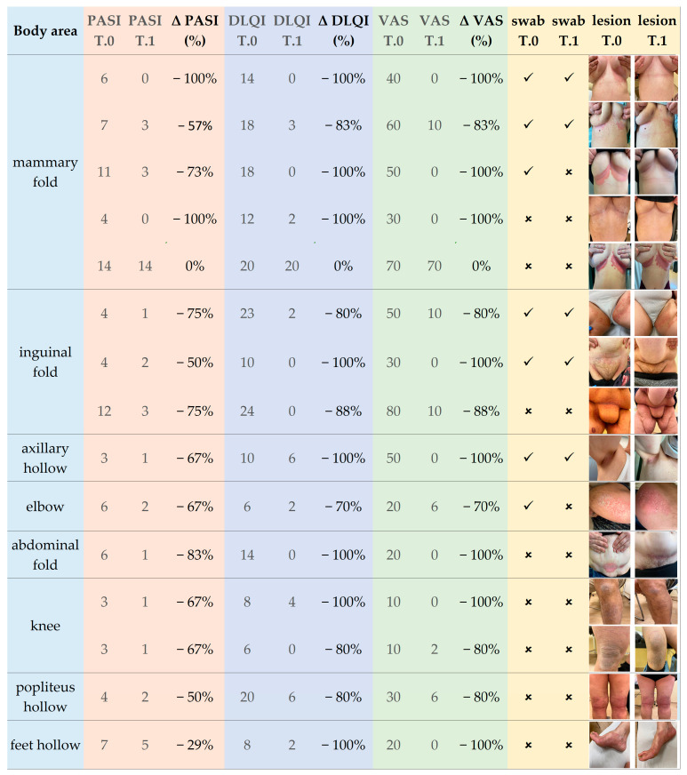
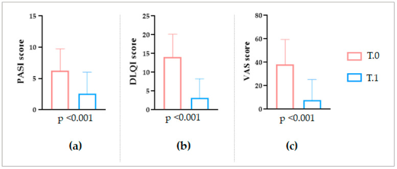
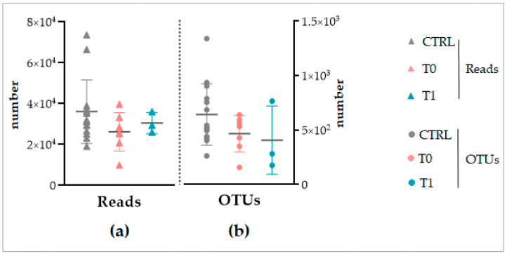
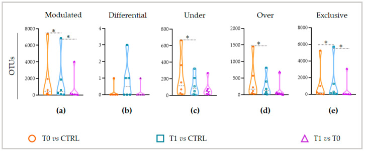
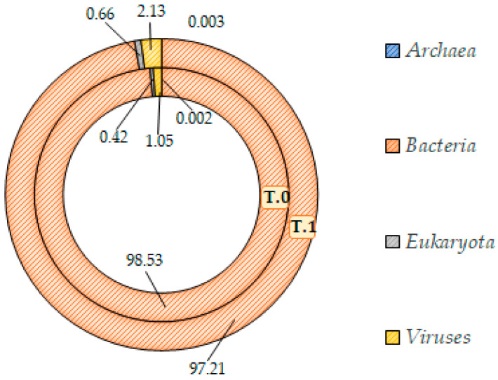
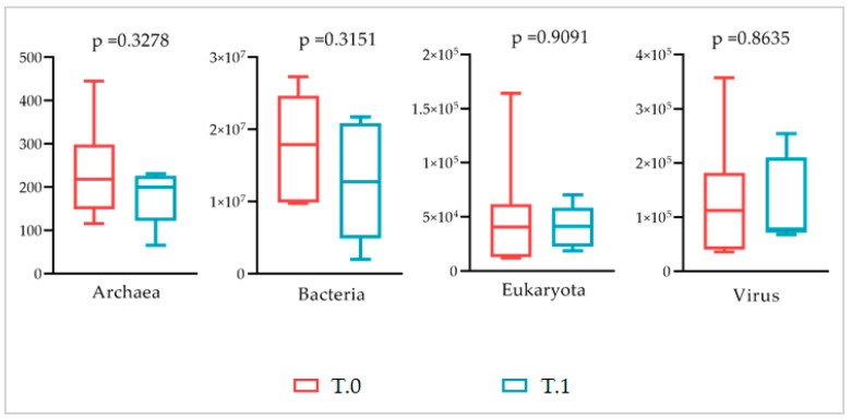
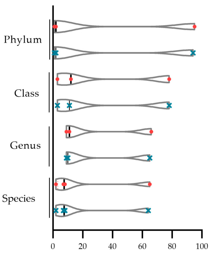
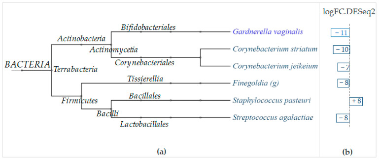
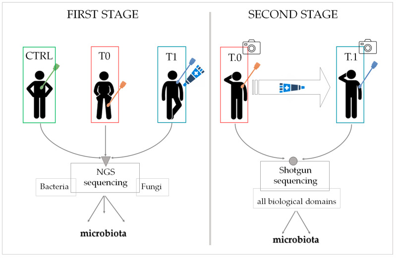
(1) Inverse psoriasis (IP), also known as intertriginous, typically affects the groin, armpits, navel, intergluteal fissure, and external genitalia. Skin lesions are erythematous plaques of inflammatory nature, smooth, well-delimited, non-scaly, and non-infiltrated. Lesions may be accompanied by itching, pain, or burning sensation. The aim of this study is both to investigate the modulation of the skin microbiota induced by IP and, on the other hand, to test the effectiveness of the new biotechnological product LimpiAL 2.5%. (2) Patients affected by IP were recruited in a private practice and treated for 4 weeks with LimpiAL 2.5% exclusively. The clinical effects on the lesion skin were evaluated, and the skin microbiotas before and after treatment were compared. (3) The clinical outcomes reveled a significant beneficial effect of the tested product. At the same time, LimpiAL increased the biological diversity of the skin microbiota and exerted a significant decrease of some Corynebacterium species, and the increase of some Staphylococcus species. (4) Together, the clinical outcomes and the microbiota analysis suggest that LimpiAL treatment improves the skin condition of affected patients, basically restoring the eubiosis conditions of the affected sites and modulating the bacterial composition of the resident microbiota.
Keywords: Actinobacteria; Corynebacterium; Firmicutes; Staphylococcus; fold; intertriginous; inverse; microbiota; psoriasis; treatment.
Conflict of interest statement
All the authors firmly declare they avoided circumstances that might affect their judgement or impartiality. Therefore, all the authors declare that there are no conflict of interest. The funder financed the material and analysis costs. Instead, it had no role in the design of the study; in the collection, analyses, or interpretation of data; in the writing of the manuscript; or in the decision to publish the results.
Figures
References
-
- Khosravi H., Siegel M.P., Van Voorhees A.S., Merola J.F. Treatment of Inverse/Intertriginous Psoriasis: Updated Guidelines from the Medical Board of the National Psoriasis Foundation. J. Drugs Dermatol. 2017;16:760–766. - PubMed
MeSH terms
LinkOut - more resources
Full Text Sources
Medical
