. 2023 Mar 28;24(7):6341.
doi: 10.3390/ijms24076341.

Mir-302a/TWF1 Axis Impairs the Myogenic Differentiation of Progenitor Cells through F-Actin-Mediated YAP1 Activation

Affiliations

Mir-302a/TWF1 Axis Impairs the Myogenic Differentiation of Progenitor Cells through F-Actin-Mediated YAP1 Activation

Mai Thi Nguyen et al. Int J Mol Sci. .

Abstract

Actin cytoskeleton dynamics have been found to regulate myogenesis in various progenitor cells, and twinfilin-1 (TWF1), an actin-depolymerizing factor, plays a vital role in actin dynamics and myoblast differentiation. Nevertheless, the molecular mechanisms underlying the epigenetic regulation and biological significance of TWF1 in obesity and muscle wasting have not been explored. Here, we investigated the roles of miR-302a in TWF1 expression, actin filament modulation, proliferation, and myogenic differentiation in C2C12 progenitor cells. Palmitic acid, the most prevalent saturated fatty acid (SFA) in the diet, decreased the expression of TWF1 and impeded myogenic differentiation while increasing the miR-302a levels in C2C12 myoblasts. Interestingly, miR-302a inhibited TWF1 expression directly by targeting its 3'UTR. Furthermore, ectopic expression of miR-302a promoted cell cycle progression and proliferation by increasing the filamentous actin (F-actin) accumulation, which facilitated the nuclear translocation of Yes-associated protein 1 (YAP1). Consequently, by suppressing the expressions of myogenic factors, i.e., MyoD, MyoG, and MyHC, miR-302a impaired myoblast differentiation. Hence, this study demonstrated that SFA-inducible miR-302a suppresses TWF1 expression epigenetically and impairs myogenic differentiation by facilitating myoblast proliferation via F-actin-mediated YAP1 activation.

Keywords: YAP1; differentiation; miR-302a; myogenesis; proliferation; twinfilin-1.


Conflict of interest statement

The authors declare no conflict of interest.

Figures

Figure 1
Figure 1
PA impairs myogenic differentiation and modulates TWF1 and miR-302a expression. C2C12 myoblasts were treated with PA (100 μM) or a vehicle for 24 h and differentiated for 5 days. (A) Immunofluorescence staining for MyHC (MF20, green) and nucleus (Hoechst 34452, blue) on differentiation day 5. Scale bar: 50 μm. (B) Myogenic differentiation was analyzed using MyHC-positivity areas, differentiation and fusion indices as described in the Materials and Methods. (C,D) The expression levels of myogenic factors (MyoD, MyoG, and MyHC) and TWF1 were determined by immunoblots on differentiation day 3 and normalized versus β-actin by densitometry. (E) The expression levels of miR-302a were analyzed by RT-qPCR after 24 h of PA treatment and normalized versus U6. Results are expressed as relative ratios versus vehicle controls and presented as means ± standard errors (n > 3); **, p < 0.01; ***, p < 0.001 vs. controls.
Figure 2
Figure 2
MiR-302a suppresses TWF1 expression by directly binding to TWF1 3′UTR. (A) Schematic diagram showing the potential miR-302a binding site on the 3’UTR of TWF1. (B) Sequences of the miR-302a binding sites of wild-type (TWF1wt) and mutant (TWF1mut) TWF1 3’UTR. (C) The dual-luciferase reporter assay was conducted 24 h after transfection in C2C12 cells. (D) Representative immunoblots of TWF1 in C2C12 cells 24 h after transfection with scRNA or miR-302a mimic. Immunoblot results are expressed as relative ratios versus scRNA controls and presented as means ± standard errors (n > 3); **, p < 0.01; ***, p < 0.001 vs. scRNA. ns: no significance.
Figure 3
Figure 3
MiR-302a promotes F-actin formation and the nuclear localization of YAP1. C2C12 cells were transfected with scRNA, siTWF1, miR-302a mimic, or antimiR-302 for 24 h before analysis. (A,B) F-actin was stained with FITC-phalloidin (green). Nuclei were counterstained with Hoechst 34452 (blue). Scale bar: 25 μm. (C,D) Immunoblots of YAP1 and phosphorylated YAP1 (pYAP1) in the nuclear and cytoplasmic fractions. α-Tubulin and Lamin B were applied as cytoplasmic and nuclear fraction markers, respectively. Immunoblot results are expressed as relative ratios versus Lamin B or α-Tubulin and presented as means ± standard errors (n > 3); ***, p < 0.001 vs. scRNA. ns: no significance.
Figure 4
Figure 4
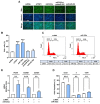
MiR-302a promotes myoblast proliferation. C2C12 cells were transfected with scRNA, siTWF1, miR-302a mimic, or antimiR-302 for 24 h before analysis. (A) EdU incorporation analysis. Cells undergoing DNA replication were labeled with EdU (green) and nuclei were counterstained with Hoechst 34452 (blue). Scale bar: 50 μm. (B) Proportions of EdU-positive cells were determined using ImageJ software. (C,D) Representative flow cytometry results. The scatter plots of cell cycle images and cell cycle phase proportions were obtained after 24 h of transfection. (E) Relative expression levels of CCNB1, PCNA, and CCND1 were determined via RT-qPCR and normalized versus U6. qRT-PCR results are expressed as relative ratios versus controls (scRNA) and presented as means ± standard errors (n > 3); **, p < 0.01; ***, p < 0.001 vs. scRNA.
Figure 5
Figure 5
MiR-302a inhibits the expressions of myogenic factors and TWF1. C2C12 cells were transfected with scRNA, siTWF1, miR-302a mimic, or antimiR-302 and differentiated for 3 days. (A) Representative immunoblots of MyoD, MyoG, MyHC, and TWF1 with β-actin are shown. (B) Quantitative analysis of TWF1 and myogenic factor (MyHC, MyoD, and MyoG) protein levels. Intensities of immunoblots were normalized versus β-actin via densitometry. Values are relative ratios versus controls (scRNA). Results are presented as means ± standard errors (n > 3); ***, p < 0.001 vs. scRNA.
Figure 6
Figure 6
MiR-302a impairs myogenic differentiation. C2C12 cells were transfected with scRNA, siTWF1, miR-302a mimic, or antimiR-302 and differentiated for 5 days. (A) Immunofluorescence staining for MyHC (MF20, green) and nucleus (Hoechst 34452, blue) on differentiation day 5. Scale bar: 50 μm. (B) Myogenic differentiation was analyzed using MyHC-positivity areas, differentiation and fusion indices, and myotube widths. Values are relative ratios versus controls (scRNA). Results are presented as means ± standard errors (n > 3); ***, p < 0.001 vs. scRNA.

Similar articles

References

    1. Mukund K., Subramaniam S. Skeletal muscle: A review of molecular structure and function, in health and disease. Wiley Interdiscip. Rev. Syst. Biol. Med. 2020;12:e1462. doi: 10.1002/wsbm.1462. - DOI - PMC - PubMed
    1. Yusuf F., Brand-Saberi B. Myogenesis and muscle regeneration. Histochem. Cell Biol. 2012;138:187–199. doi: 10.1007/s00418-012-0972-x. - DOI - PubMed
    1. Chal J., Pourquie O. Making muscle: Skeletal myogenesis in vivo and in vitro. Development. 2017;144:2104–2122. doi: 10.1242/dev.151035. - DOI - PubMed
    1. Sartori R., Romanello V., Sandri M. Mechanisms of muscle atrophy and hypertrophy: Implications in health and disease. Nat. Commun. 2021;12:330. doi: 10.1038/s41467-020-20123-1. - DOI - PMC - PubMed
    1. Akhmedov D., Berdeaux R. The effects of obesity on skeletal muscle regeneration. Front. Physiol. 2013;4:371. doi: 10.3389/fphys.2013.00371. - DOI - PMC - PubMed

MeSH terms