. 2023 Mar 28;24(7):6353.
doi: 10.3390/ijms24076353.

Dental Follicle Stem Cells Promote Periodontal Regeneration through Periostin-Mediated Macrophage Infiltration and Reprogramming in an Inflammatory Microenvironment

Affiliations

Dental Follicle Stem Cells Promote Periodontal Regeneration through Periostin-Mediated Macrophage Infiltration and Reprogramming in an Inflammatory Microenvironment

Xiuqun Wei et al. Int J Mol Sci. .

Abstract

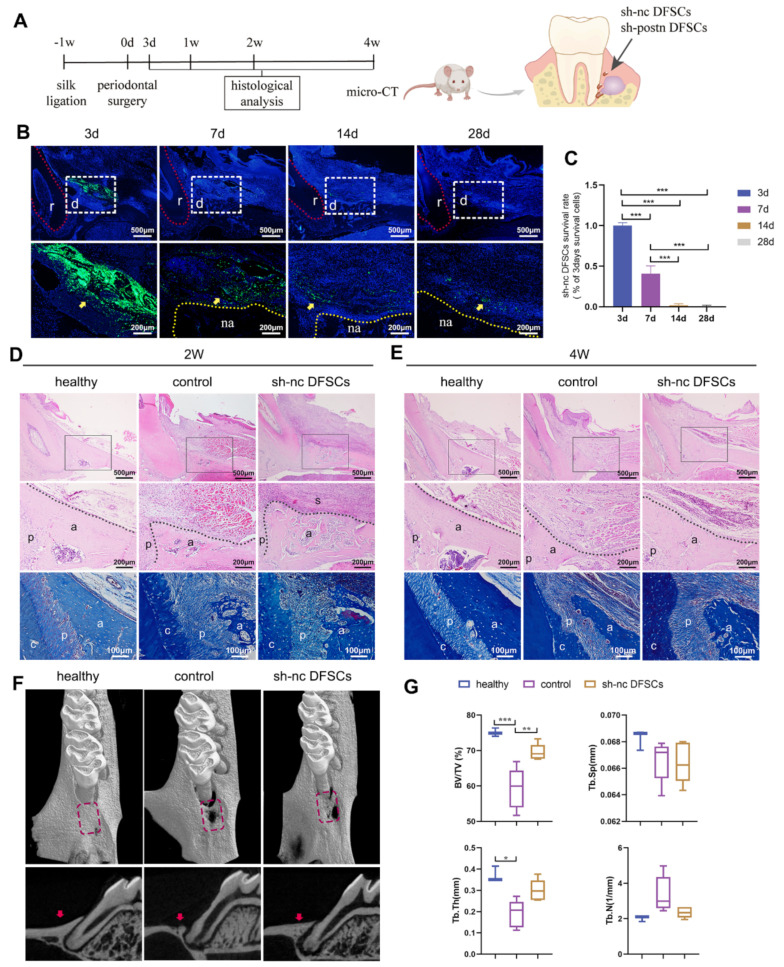
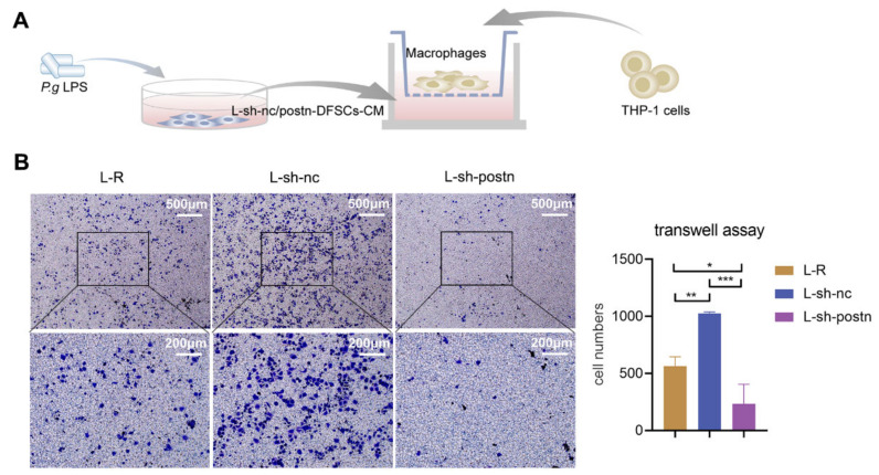
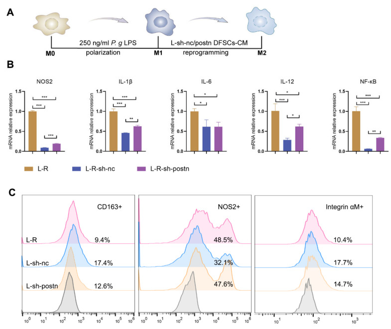
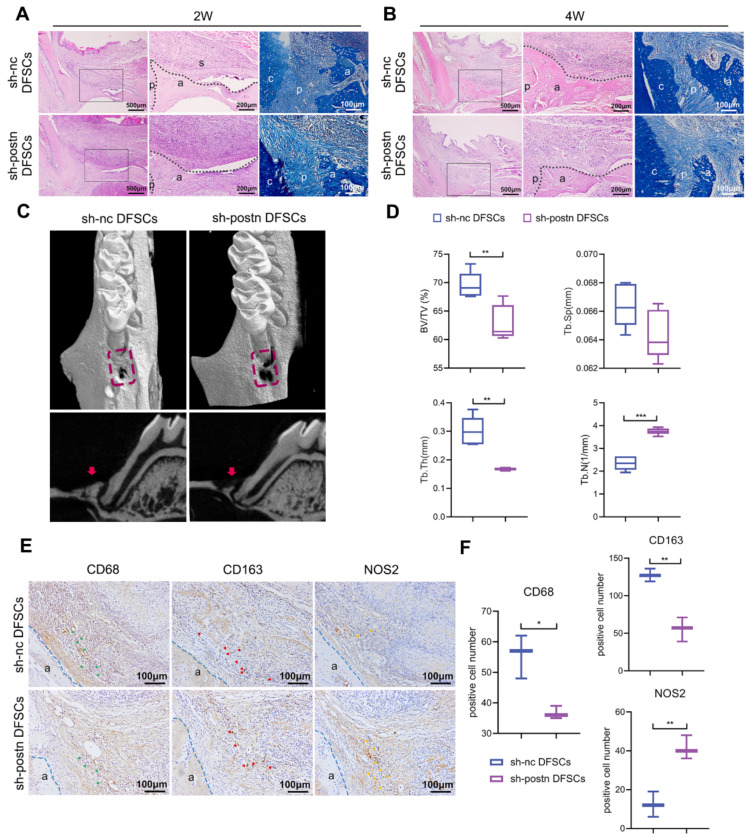
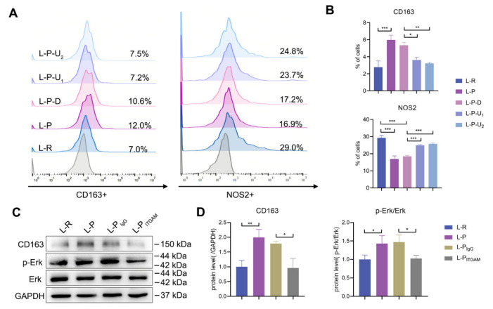
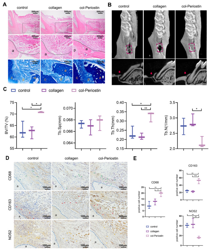
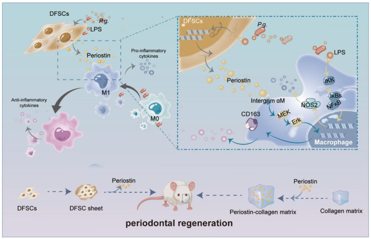
Dental follicle stem cells (DFSCs) have been verified to promote periodontal regeneration in an inflammatory microenvironment. When coping with inflammatory stimulation, DFSCs highly express periostin, a bioactive molecule closely related to periodontal homeostasis. It is worth exploring whether and how periostin plays a role in the promotion of periodontal regeneration by DFSCs. By tracking the fate of DFSCs, it was found that DFSCs significantly contributed to periodontal regeneration in rat periodontal defects while they had a low survival rate. They highly expressed periostin and improved the immune microenvironment in the defect area, especially via the recruitment and reprogramming of macrophages. Silencing periostin attenuated the effects of DFSCs in promoting periodontal regeneration and regulating macrophages. Recombinant human periostin (rhPeriostin) could not only directly promote macrophage reprogramming through the integrin αM/phosphorylated extracellular signal-regulated kinase (p-Erk)/Erk signaling pathway, but it also exhibited the potential to promote periodontal regeneration in rats when loaded in a collagen matrix. These results indicated that periostin is actively involved in the process by which DFSCs promote periodontal regeneration through the regulation of macrophages and is a promising molecular agent to promote periodontal regeneration. This study provides new insight into the mechanism by which DFSCs promote periodontal regeneration and suggests a new approach for periodontal regeneration therapy.

Keywords: dental follicle stem cells; macrophage reprogramming; periodontal regeneration; periostin.


Conflict of interest statement

The authors declare no conflict of interest.

Figures

Figure 1
Figure 1
The therapeutic effect and survival rate of DFSCs. (A) Schematic illustration of animal model. Fluorescence images (B) and related analysis (C) of DFSC survival. The red dotted line indicates the root. The data were analyzed by one-way analysis of variance (ANOVA) and Bonferroni post hoc test (n = 3). The white dashed box indicates the defect area. The yellow dotted line indicates the alveolar bone. The yellow arrow indicates DFSCs. r: root; d: defect area; na: newly formed alveolar bone. HE and Masson’s trichrome (MT) staining at 2 weeks (D) and 4 weeks (E). Middle panel represents a magnified view of the black box in the upper panel. The black dotted line shows the newly formed alveolar bone. a: alveolar bone; c: cementum; p: periodontal ligament, s: DFSC sheet. Micro-computed tomography (micro-CT) images (F) and quantitative assessment (G) of the defect area at 4 weeks (n = 5). The data of Tb.Sp and Tb.N were analyzed by one-way ANOVA and Bonferroni post hoc test. The data of Tb.Th did not follow normal distribution and were analyzed by Kruskal-Wallis test. The rectangular area and red arrow indicate the defect area in the 3D and sagittal views, respectively. * p < 0.05; ** p < 0.01; *** p < 0.001. Error bars represent means ± SD.
Figure 2
Figure 2
DFSCs improved the immune microenvironment and highly expressed periostin. (A) HE staining of the defect areas of the control and sh-nc DFSCs groups at 3 days after transplantation. The same area is shown in the healthy group. Right panel represents a magnified view of the black box in the left panel. The immunochemistry images (B) and quantification analysis (C) of the CD68+, CD163+, and NOS2+ cells on day 14 after surgery. The data were analyzed by one-way analysis of variance (ANOVA) and Bonferroni post hoc test (n = 3). The blue dotted line indicates the outline of the alveolar bone. a: alveolar bone; p: periodontal ligament. (D) Representative images of immunofluorescence staining against periostin in the defect area at 3 and 7 days. Data from 3d and 7d were analyzed independently by one-way analysis of variance (ANOVA) and Bonferroni post hoc test (n = 3). Data from the same treatment group at different time points were analyzed by unpaired two-tailed Student’s t-test (n = 3).; ** p < 0.01; *** p < 0.001. Error bars represent means ± SD.
Figure 3
Figure 3
Silencing periostin in DFSCs prevented macrophage migration. (A) Schematic diagram to validate the effect of knocking down periostin on DFSC-promoted macrophage migration. (B) Representative images and quantitative analysis of macrophage migration under the DFSCs-CM. The data were analyzed by one-way ANOVA and Bonferroni post hoc test (n = 3). Images were taken at either 4× (upper row) or 10× (lower row) magnification. * p < 0.05; ** p < 0.01; *** p < 0.001. Error bars represent means ± SD.
Figure 4
Figure 4
Silencing periostin inhibited the reprogramming of macrophages to the M2 type by DFSCs. (A) Diagram of the strategy for validating that periostin affects the ability of DFSCs to reprogram M1 to M2 macrophages. (B) qRT-PCR for M1 marker and pro-inflammatory factors in macrophages. The data were analyzed by one-way ANOVA and Bonferroni post hoc test (n = 3). (C) Flow cytometric analysis for surface marker expression on macrophages. * p < 0.05; ** p < 0.01; *** p < 0.001. Error bars represent means ± SD.
Figure 5
Figure 5
Silencing periostin affected the ability of DFSCs to promote periodontal regeneration. HE and MT staining of periodontal regeneration at 2 weeks (A) and 4 weeks (B). Middle panel represents a magnified view indicated by the black box in the left panel. The black dotted line shows the edge of the newly formed alveolar bone. a: alveolar bone; c: cementum; p: periodontal ligament; s: DFSCs sheet. Micro-CT images (C) and quantitative assessment (D) of the defect area at 4 weeks (n = 5). The red dotted box indicates the defect area and the red arrow indicates regenerated alveolar bone. The data of BV/TV, Tb.Sp, and Tb.N were analyzed by unpaired two-tailed Student’s t test. Unpaired two-tailed t tests with Welch correction were used for Tb.Th. The immunochemistry images (E) and quantification analysis (F) of the CD68+, CD163+, and NOS2+ cells on day 14 after surgery. green arrows: CD68+ cells; red arrows: CD163+ cells; yellow arrows: NOS2+ cells. The data were analyzed by unpaired two-tailed Student’s t test (n = 3). The blue dotted line indicates the outline of the alveolar bone. a: alveolar bone; p: periodontal ligament. * p < 0.05; ** p < 0.01; *** p < 0.001. Error bars represent means ± SD.
Figure 6
Figure 6
Periostin reprogrammed macrophages partly through integrin αM/p-Erk/Erk signaling. (A,B) Flow cytometric analysis for pro- and anti-inflammatory marker expression in macrophages after periostin stimulation with or without Erk inhibitor. The data were analyzed by one-way ANOVA and Bonferroni post hoc test (n = 3). L-R: group treated with RPMI1640 alone; L-P: group treated with rhPeriostin; L-P-D: group treated with rhPeriostin and DMSO; L-P-U1: group treated with rhPeriostin and 10 μg/mL U0126; L-P-U2: group treated with rhPeriostin and 20 μg/mL U0126. (C,D) Western blot analysis of CD163 and phosphorylation of ERK in macrophages after periostin stimulation with or without anti-ITGAM antibodies. IgG isotype control or anti-ITGAM antibodies were added to examine the role of integrin αM in signaling. The data were analyzed by one-way ANOVA and Bonferroni post hoc test (n = 3). * p < 0.05; ** p < 0.01; *** p < 0.001. Error bars represent means ± SD.
Figure 7
Figure 7
RhPeriostin promoted periodontal tissue regeneration. (A) HE and MT staining of periodontal regeneration at 4 weeks after periostin treatment. Images shown are representative images. Middle panel represents a magnified view of the black box in the upper panel. The black dotted line shows the edge of the newly formed alveolar bone. a: alveolar bone; c: cementum; p: periodontal ligament. Micro-CT images (B) and quantitative assessment (C) of the defect area at 4 weeks (n = 3). The red dotted box indicates the defect area, and the red arrow indicates regenerated alveolar bone. The data of BV/TV, Tb.Sp, Tb.N, and Tb.Th were analyzed by one-way ANOVA and Bonferroni post hoc test. The immunochemistry images (D) and quantification analysis (E) of the CD68+, CD163+, and NOS2+ cells on day 28 after surgery. green arrows: CD68+ cells; red arrows: CD163+ cells; yellow arrows: NOS2+ cells. The data were analyzed by one-way ANOVA and Bonferroni post hoc test (n = 3). The blue dotted line indicates the outline of the alveolar bone. a: alveolar bone. * p < 0.05; ** p < 0.01; *** p < 0.001. Error bars represent means ± SD.
Figure 8
Figure 8
Schematic diagram of the role and mechanism of periostin in the promotion of periodontal regeneration by DFSCs. Periostin is actively involved in DFSC-mediated periodontal regeneration through regulating macrophages via integrin αM/p-Erk/Erk signaling and is a promising molecular agent to promote periodontal regeneration.

Similar articles

Cited by

References

    1. Tonetti M.S., Greenwell H., Kornman K.S. Staging and grading of periodontitis: Framework and proposal of a new classification and case definition. J. Periodontol. 2018;89:S159–S172. doi: 10.1002/JPER.18-0006. - DOI - PubMed
    1. Nuñez J., Vignoletti F., Caffesse R.G., Sanz M. Cellular therapy in periodontal regeneration. Periodontology 2000. 2019;79:107–116. doi: 10.1111/prd.12250. - DOI - PubMed
    1. Yan N., Hu B., Xu J., Cai R., Liu Z., Fu D., Huo B., Liu Z., Zhao Y., Chen C., et al. Stem cell Janus patch for periodontal regeneration. Nano Today. 2022;42:101336. doi: 10.1016/j.nantod.2021.101336. - DOI
    1. Li Q., Yang G., Li J., Ding M., Zhou N., Dong H., Mou Y. Stem cell therapies for periodontal tissue regeneration: A network meta-analysis of preclinical studies. Stem Cell Res. Ther. 2020;11:427. doi: 10.1186/s13287-020-01938-7. - DOI - PMC - PubMed
    1. Wang P., Wang W., Geng T., Liu Y., Zhu S., Liu Z., Yuan C. EphrinB2 regulates osteogenic differentiation of periodontal ligament stem cells and alveolar bone defect regeneration in beagles. J. Tissue Eng. 2019;10:2041731419894361. doi: 10.1177/2041731419894361. - DOI - PMC - PubMed