Review
. 2023 Mar 28;24(7):6357.
doi: 10.3390/ijms24076357.

3D Bioprinting for Next-Generation Personalized Medicine

Affiliations
Review

3D Bioprinting for Next-Generation Personalized Medicine

Ethan Hau Yin Lam et al. Int J Mol Sci. .

Abstract

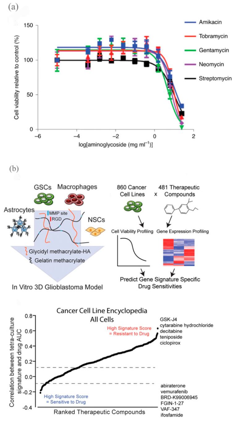
In the past decade, immense progress has been made in advancing personalized medicine to effectively address patient-specific disease complexities in order to develop individualized treatment strategies. In particular, the emergence of 3D bioprinting for in vitro models of tissue and organ engineering presents novel opportunities to improve personalized medicine. However, the existing bioprinted constructs are not yet able to fulfill the ultimate goal: an anatomically realistic organ with mature biological functions. Current bioprinting approaches have technical challenges in terms of precise cell deposition, effective differentiation, proper vascularization, and innervation. This review introduces the principles and realizations of bioprinting with a strong focus on the predominant techniques, including extrusion printing and digital light processing (DLP). We further discussed the applications of bioprinted constructs, including the engraftment of stem cells as personalized implants for regenerative medicine and in vitro high-throughput drug development models for drug discovery. While no one-size-fits-all approach to bioprinting has emerged, the rapid progress and promising results of preliminary studies have demonstrated that bioprinting could serve as an empowering technology to resolve critical challenges in personalized medicine.

Keywords: biomaterial; bioprinting; drug discovery; personalized medicine; precision medicine; regenerative medicine; stem cell.


Conflict of interest statement

The authors declare no conflict of interest.

Figures

Figure 1
Figure 1
A brief timeline of major scientific discoveries and events [5,6,7,8,9,10,11,12,13,14,15,16,17].
Figure 2
Figure 2
Four main bioprinting methods. (a) Inkjet Bioprinting. (b) Extrusion Bioprinting. (c) Laser-Assisted Bioprinting. (d) Stereolithography Bioprinting were reprinted with permission from Ref. [14]. 2020, Yu. This figure is modified, and obtained from an open-access journal article.
Figure 3
Figure 3
A schematic of factors under consideration for the bioprinting of stem cells.
Figure 4
Figure 4
Cellular extrusion bioprinting improved nephron formation in kidney organoid, as compared to manual generation. (A) A schematic of the protocol for manual versus bioprinted kidney organoid formation (R40, R0). R40 and R0 were generated from 1.1 × 10⁵ differentiated iPSC (MAFBmTAGBFP2GATA3mCherry) cells, and manual organoids from 2.3 × 10⁵ cells. (B) Comparison of MAFB reporter area in manual and bioprinted kidney organoids. Larger area was observed in R40 organoids, suggesting greater nephron number formation. Bars indicate mean. R40-Man, p = 2.1 × 10⁻⁵, R40-R0, p = 2 × 10⁻¹⁶. (C) Uniform formation of nephron structures in the bioprinted kidney organoid patch, analyzed by brightfield imaging. (D) Nephrons showed expression of markers of proximal tubules (LTL (left panel; green) and HNF4A (right panel; red)), podocytes (mTagBFP2 (left panel; blue)), nephron epithelium (EPCAM (left panel; red)), distal tubule/loop of Henle TAL (SLC12A1 (right panel; green)), surrounded by interstitial endothelial cells (SOX17 (right panel; grey)). Analyzed by confocal immunofluorescence imaging. (E) Patch organoid was generated from proximal tubule-specific iPSC reporter line (where yellow fluorescent protein (YFP) was inserted under the control of the HNF4A promoter), following incubation in TRITC-albumin substrate. Live confocal imaging shows uptake of TRITC-albumin (red) into YFP-positive proximal tubules (yellow). Small panels below show higher magnification of the outlined areas, with and without phase-contrast overlays. Scale bars = 100 μm. Reproduced with permission from [68], copyright 2020 Springer Nature Ltd.
Figure 5
Figure 5
A scaffold-free approach to heart regeneration through printing cardiac spheroids containing human-induced pluripotent stem-cell-derived cardiomyocytes. (a) Surgical procedures of mouse myocardial infarction model. (b) Patch group: one cardiac patch (CP) and a natural omentum patch (OP) were sutured over the site of infarction. (c) One OP was sutured over the site of infarction. (d) Schematics of experimental setup. (e,f) vascularization of the infarcted area. Patch group vs. control group. Green: positive staining of the endothelial cell. Scale bar = 100 μm. (g) Cardiac output (mL/min). Patch group vs. control group. (CO: 104.6 ± 45.5 vs. 68.6 ± 16.4, p = 0.1). Reproduced with permission from [70], copyright 2019 John Wiley & Sons, Ltd.
Figure 6
Figure 6
Bioprinted cardiovascular and liver models for drug development. (a) Schematic diagram of cardiac tissue model design with stress sensors (scale bars are 10 mm) and verapamil and isoproterenol dose-response plots below. Adapted with permission from [85]. 2016, Nature Publishing Group. (b) DLP-bioprinted hepatic model on left, fluorescence and bright field images of 3D-printed construct on top-right, and CYP induction plots on lower-right with asterisks indicating statistical significance with threshold of p < 0.05 (scale bars are 500 mm). Adapted with permission from [86]. 2015, S. Chen.
Figure 7
Figure 7
Bioprinted kidney and glioblastoma models for drug development. (a) Application of 96-well bioprinted organoids for testing viability in response to aminoglycoside antibiotics. The curves represent a non-linear fit for each compound, with n = 19 (amikacin), n = 24 (tobramycin), n = 30 (gentamycin), n = 30 (neomycin), n = 22 (streptomycin). Adapted from [68]. 2020, Nature Publishing Group. (b) Development of 3D glioblastoma model involving glioblastoma stem cells (GSCs), macrophages, astrocytes, and neural stem cells (NSCs) on left, methodology of evaluating drug sensitivity based on 3D tetra-culture gene expression signature from Cancer Cell Line Encyclopedia (CCLE) and the Cancer Therapeutic Response Platform (CTRP) datasets in middle, therapeutic efficacy prediction of drugs in CTRP dataset cancer cells using differentially expressed genes (as determined by RNA-sequencing) between 3D tetra-culture model and GSCs grown in sphere culture. Adapted from [92]. 2020, Nature Publishing Group.

References

    1. Gorshkov K., Chen C.Z., Marshall R.E., Mihatov N., Choi Y., Nguyen D.-T., Southall N., Chen K., Park J.K., Zheng W. Advancing Precision Medicine with Personalized Drug Screening. Drug Discov. Today. 2019;24:272–278. doi: 10.1016/j.drudis.2018.08.010. - DOI - PMC - PubMed
    1. Ho D., Quake S.R., McCabe E.R.B., Chng W.J., Chow E.K., Ding X., Gelb B.D., Ginsburg G.S., Hassenstab J., Ho C.-M., et al. Enabling Technologies for Personalized and Precision Medicine. Trends Biotechnol. 2020;38:497–518. doi: 10.1016/j.tibtech.2019.12.021. - DOI - PMC - PubMed
    1. Li Y., Nieuwenhuis L.M., Keating B.J., Festen E.A.M., de Meijer V.E. The Impact of Donor and Recipient Genetic Variation on Outcomes After Solid Organ Transplantation: A Scoping Review and Future Perspectives. Transplantation. 2022;106:1548–1557. doi: 10.1097/TP.0000000000004042. - DOI - PMC - PubMed
    1. Mathur S., Sutton J. Personalized Medicine Could Transform Healthcare. Biomed. Rep. 2017;7:3–5. doi: 10.3892/br.2017.922. - DOI - PMC - PubMed
    1. Takahashi K., Tanabe K., Ohnuki M., Narita M., Ichisaka T., Tomoda K., Yamanaka S. Induction of Pluripotent Stem Cells from Adult Human Fibroblasts by Defined Factors. Cell. 2007;131:861–872. doi: 10.1016/j.cell.2007.11.019. - DOI - PubMed

LinkOut - more resources