. 2023 Mar 28;24(7):6367.
doi: 10.3390/ijms24076367.

Investigation of the Entry Pathway and Molecular Nature of σ1 Receptor Ligands

Affiliations

Investigation of the Entry Pathway and Molecular Nature of σ1 Receptor Ligands

Gianmarco Pascarella et al. Int J Mol Sci. .

Abstract

The σ1 receptor (σ1-R) is an enigmatic endoplasmic reticulum resident transmembrane protein implicated in a variety of central nervous system disorders and whose agonists have neuroprotective activity. In spite of σ1-R's physio-pathological and pharmacological importance, two of the most important features required to fully understand σ1-R function, namely the receptor endogenous ligand(s) and the molecular mechanism of ligand access to the binding site, have not yet been unequivocally determined. In this work, we performed molecular dynamics (MD) simulations to help clarify the potential route of access of ligand(s) to the σ1-R binding site, on which discordant results had been reported in the literature. Further, we combined computational and experimental procedures (i.e., virtual screening (VS), electron density map fitting and fluorescence titration experiments) to provide indications about the nature of σ1-R endogenous ligand(s). Our MD simulations on human σ1-R suggested that ligands access the binding site through a cavity that opens on the protein surface in contact with the membrane, in agreement with previous experimental studies on σ1-R from Xenopus laevis. Additionally, steroids were found to be among the preferred σ1-R ligands predicted by VS, and 16,17-didehydroprogesterone was shown by fluorescence titration to bind human σ1-R, with significantly higher affinity than the prototypic σ1-R ligand pridopidine in the same essay. These results support the hypothesis that steroids are among the most important physiological σ1-R ligands.

Keywords: Huntington’s disease; fluorescence titration; molecular dynamics; virtual screening; σ1 receptor.


Conflict of interest statement

The authors declare no conflict of interest.

Figures
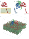
Figure 1
Figure 1
Structure of Hsσ1-R protein. (A) A single monomer is shown as ribbon. The position of the monomer with respect to the membrane region of the protein is highlighted. (B) The homotrimer is shown as ribbon. The three monomers (MA, MB and MC) are colored blue, red and green, respectively. (C) Simulated system within the simulation box. Membrane atoms are shown as spheres and colored by atom type: C, O, N, P and H are green, red, blue, orange and white, respectively.
Figure 2
Figure 2
Conformational changes occurring in the Hsσ1-R protein along the simulated trajectory. (A) The distances between the mass centers of the backbone atoms of residues 118–121 and 180–188 for the three monomers (namely, MA, MB and MC) are reported as a function of time. (B,C) Cartoon representation of the Hsσ1-R protein viewed from the membrane side at 0 ns (B) and 1500 ns (C) of the MD simulation. The three monomers are colored blue (M1), red (M2) and green (M3). The surface of residues 118–121 and 180–188 of the three monomers is also shown and colored grey and yellow, respectively. (D,E) Cartoon representation of the Hsσ1-R protein C-terminal domains, external to the membrane, at 0 ns (D) and 1500 ns (E) of the MD simulation. The surface of residues 122–126 and 171–176 of M2 (red) is also shown and colored grey and yellow, respectively, highlighting the opening of the Hsσ1-R binding site that occurs along the simulated trajectory.
Figure 3
Figure 3
Time evolution of salt bridges between residues R175, E102 and E123 along the MD simulation. In the left panel, the minimum distances calculated between R175 and E102 (black) and between R175 and E123 (orange) are reported as a function of the simulated time for the three monomers, namely M1 (top panel), M2 (middle panel) and M3 (bottom panel). Right panel: cartoon representation of M2 in the starting conformation of the MD simulation, which is virtually identical to the crystallographic conformation. Zoomed-in inset: residues R175, E102 and E123 are shown as sticks and coloured by atom type: C, cyan; N, blue; O, red; H, white.
Figure 4
Figure 4
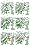
Fitting of selected compounds into the electron density within the ligand binding site of Xlσ1-R. The protein is shown as ribbon and colored green. The side-chains of residues surrounding the ligand binding site are shown as sticks and colored by atom-type: C, N, O and S atoms are green, blue, red and yellow, respectively. The structures in coordinate files 7W2B and 7W2E are shown in panels (AF) and (GL), respectively. Ligands in panels (BF) and (HL) are shown as sticks and colored by atom-type in the same way as protein side-chains, except that C atoms are orange. Ligands are: ergosterol, panels (B,H); catechin, panels (C,I); 7,8-dihydropteroic acid, panels (D,J); myricetin, panels (E,K); and 3′,5′-cyclic dAMP, panels (F,L).
Figure 4
Figure 4
Fitting of selected compounds into the electron density within the ligand binding site of Xlσ1-R. The protein is shown as ribbon and colored green. The side-chains of residues surrounding the ligand binding site are shown as sticks and colored by atom-type: C, N, O and S atoms are green, blue, red and yellow, respectively. The structures in coordinate files 7W2B and 7W2E are shown in panels (AF) and (GL), respectively. Ligands in panels (BF) and (HL) are shown as sticks and colored by atom-type in the same way as protein side-chains, except that C atoms are orange. Ligands are: ergosterol, panels (B,H); catechin, panels (C,I); 7,8-dihydropteroic acid, panels (D,J); myricetin, panels (E,K); and 3′,5′-cyclic dAMP, panels (F,L).
Figure 5
Figure 5
Molecular model of the complex between Hsσ1-R and 16,17-didehydroprogesterone built by VINA. The protein is shown as ribbon and colored green. The ligand and the side-chains of residues at a distance ≤ 4.0 Å from the ligand are shown as sticks and colored by atom-type: N, O and S atoms are blue, red and yellow, respectively; C is green for the protein and white for the ligand. The only exception is V84, which was removed from the picture for clarity.
Figure 6
Figure 6
Titration of Hsσ1-R with pridopidine (left panel), iloperidone (central panel) and 16,17-didehydroprogesterone (right panel). ΔF/ΔFmax values at 340 nm are plotted as a function of ligand concentration (see Section 4). Fluorescence titration analyses were performed using Equation (2) that describes a two-site model in which both sites are independent of each other and non-cooperative. Data are from three replicate experiments. Error bars are standard deviations (SD) with n = 3.
Figure 7
Figure 7
Path of ligand access to σ1-R binding site identified by MD simulations. Monomer B of Hsσ1-R in coordinate file 5HK1 at the beginning (t = 0 ns) and at the end (t = 1500 ns) of the MD simulation is shown as ribbon and colored green and lilac, respectively. Left: orientation of the monomer with respect to the ER membrane. Right: zoom-in of the image on the left. The ligand is proposed to access σ1-R from the protein side in contact with the ER membrane, either from the luminal medium or from the membrane itself. The movement of the α4 helix and of the β3, β4-β6 and β10 strands, comprising residues 102–109, 117–137 and 168–193, respectively, is clearly visible.

Similar articles

Cited by

References

    1. Schmidt H.R., Zheng S., Gurpinar E., Koehl A., Manglik A., Kruse A.C. Crystal structure of the human σ1 receptor. Nature. 2016;532:527–530. doi: 10.1038/nature17391. - DOI - PMC - PubMed
    1. Nguyen L., Lucke-Wold B.P., Mookerjee S., Kaushal N., Matsumoto R.R. Sigma-1 Receptors and Neurodegenerative Diseases: Towards a Hypothesis of Sigma-1 Receptors as Amplifiers of Neurodegeneration and Neuroprotection. Adv. Exp. Med. Biol. 2017;964:133–152. - PMC - PubMed
    1. Fukunaga K., Shinoda Y., Tagashira H. The role of SIGMAR1 gene mutation and mitochondrial dysfunction in amyotrophic lateral sclerosis. J. Pharmacol. Sci. 2015;127:36–41. doi: 10.1016/j.jphs.2014.12.012. - DOI - PubMed
    1. Mishra A.K., Mavlyutov T., Singh D., Biener G., Yang J., Oliver J.A., Ruoho A., Raicu V. The sigma-1 receptors are present in monomeric and oligomeric forms in living cells in the presence and absence of ligands. Biochem. J. 2015;466:263–271. doi: 10.1042/BJ20141321. - DOI - PMC - PubMed
    1. Schmidt H.R., Betz R.M., Dror R.O., Kruse A.C. Structural basis for σ1 receptor ligand recognition. Nat. Struct. Mol. Biol. 2018;25:981–987. doi: 10.1038/s41594-018-0137-2. - DOI - PMC - PubMed