. 2023 Mar 29;24(7):6411.
doi: 10.3390/ijms24076411.

A Novel RT-LAMP for the Detection of Different Genotypes of Crimean-Congo Haemorrhagic Fever Virus in Patients from Spain

Affiliations

A Novel RT-LAMP for the Detection of Different Genotypes of Crimean-Congo Haemorrhagic Fever Virus in Patients from Spain

Begoña Febrer-Sendra et al. Int J Mol Sci. .

Abstract

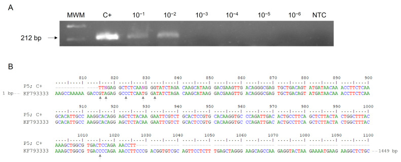
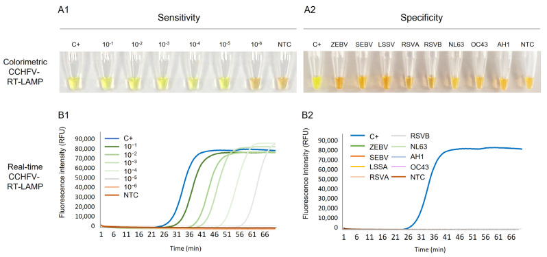
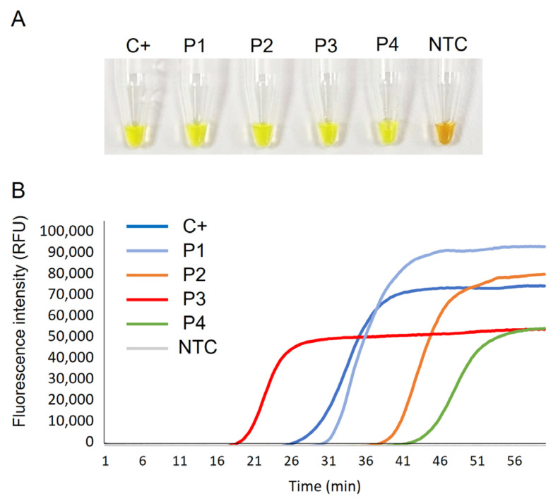
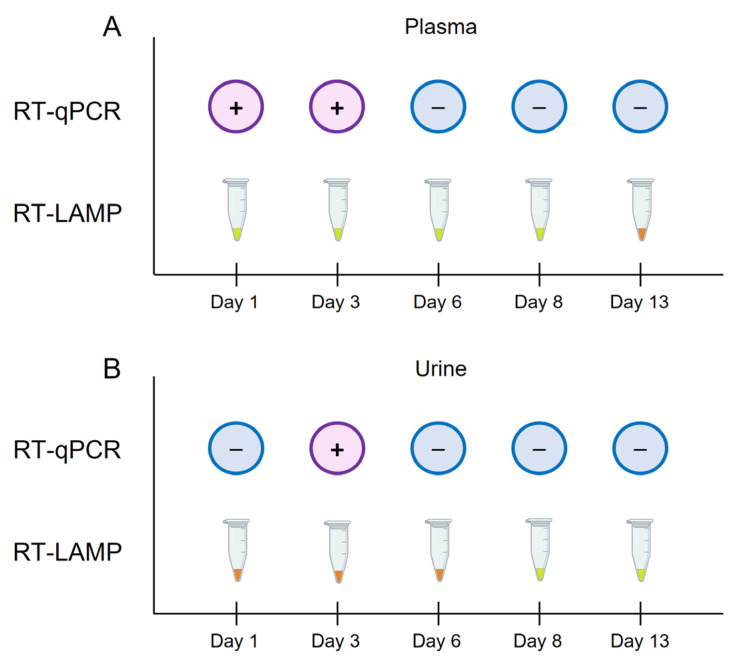
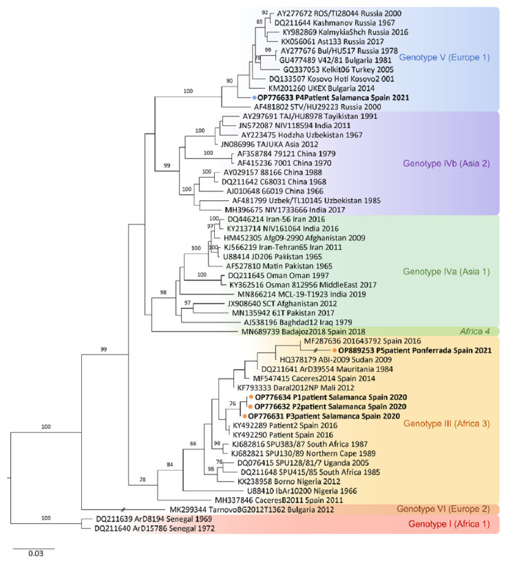
Crimean-Congo haemorrhagic fever (CCHF) is a potentially lethal tick-borne viral disease with a wide distribution. In Spain, 12 human cases of CCHF have been confirmed, with four deaths. The diagnosis of CCHF is hampered by the nonspecific symptoms, the high genetic diversity of CCHFV, and the biosafety requirements to manage the virus. RT-qPCR and serological tests are used for diagnosis with limitations. Reverse-transcription loop-mediated isothermal amplification (RT-LAMP) could be an effective alternative in the diagnosis of the disease. However, none of the few RT-LAMP assays developed to date has detected different CCHFV genotypes. Here, we designed a RT-LAMP using a degenerate primer set to compensate for the variability of the CCHFV target sequence. RT-LAMP was performed in colorimetric and real-time tests on RT-qPCR-confirmed CCHF patient samples notified in Spain in 2020 and 2021. Urine from an inpatient was analysed by RT-LAMP for the first time and compared with RT-qPCR. The amplicons obtained by RT-qPCR were sequenced and African III and European V genotypes were identified. RT-LAMP amplified both genotypes and was more sensitive than RT-qPCR in urine samples. We have developed a novel, rapid, specific, and sensitive RT-LAMP test that allows the detection of different CCHFV genotypes in clinical samples. This pan-CCHFV RT-LAMP detected viral RNA for the first time in urine samples. It can be easily performed as a single-tube isothermal colorimetric method on a portable platform in real time and without the need for expensive equipment, thus bringing molecular diagnostics closer to rural or resource-poor areas, where CCHF usually occurs.

Keywords: CCHFV; Crimean–Congo haemorrhagic fever virus; RT-LAMP; Spain; genotypes.


Conflict of interest statement

The authors declare no conflict of interest. The funders had no role in the design of the study; in the collection, analyses, or interpretation of data; in the writing of the manuscript; or in the decision to publish the results.

Figures

Figure 1
Figure 1
RT-PCR verification, sensitivity and sequencing using outer primers F3 and B3 for CCHFV RNA amplification. (A) Verification and detection limit of RT-PCR F3-B3. Lane MWM, molecular weight marker (100 bp Plus Blue DNA Ladder); lane C+, RNA isolate from patient P5 used as positive control; lanes 10−1–10−6, 10-fold serial dilutions of C+; lane NTC, non-template control (RNase-free water instead of RNA). (B) Comparison of 212 bp amplicon obtained (P5; C+) with partial sequence of Daral 2012 strain (GenBank KF793333). Grey arrows indicate different nucleotides in the sequence.
Figure 2
Figure 2
Establishing the RT-LAMP assay: sensitivity and specificity assessments for CCHFV RNA detection. (A1) Colorimetric RT-LAMP verification and sensitivity assessment. Lane C+, RNA-positive control (5 ng/µL); Lanes 10−1 to 10−6: 10-fold dilutions of CCHFV RNA sample used as positive control (C+); lane NTC, non-template control (ultra-pure water as template). (A2) Specificity assessment of colorimetric RT-LAMP. Lane C+, RNA-positive control; lanes ZEBV, SEBV, LSSV, RSVA, RSVB, NL63, OC43 and AH1: RNA from haemorrhagic viruses (Zaire Ebola virus, Sudan Ebola virus, Lassa virus) and other respiratory viruses (respiratory syncytial virus A, respiratory syncytial virus B, coronavirus NL63, coronavirus OC43 and influenza A H1, respectively. Lane NTC, non-template control (ultra-pure water as template). (B1). Real-time RT-LAMP verification and sensitivity assessment. Lane C+, RNA-positive control (5 ng/µL); Lanes 10−1 to 10−6: 10-fold dilutions of CCHFV RNA sample used as positive control (C+); lane NTC, non-template control (ultra-pure water as template). (B2) Specificity assessment of real-time RT-LAMP. Lane C+, RNA-positive control; lanes ZEBV, SEBV, LSSV, RSVA, RSVB, NL63, OC43 and AH1: RNA from haemorrhagic viruses (Zaire Ebola virus, Sudan Ebola virus, Lassa virus) and other respiratory viruses (respiratory syncytial virus A, respiratory syncytial virus B, coronavirus NL63, coronavirus OC43 and influenza A H1, respectively). Lane NTC, non-template control (ultra-pure water as template). RFU, relative fluorescence units.
Figure 3
Figure 3
Clinical sample testing by CCHFV-RT-LAMP. (A) Colorimetric RT-LAMP results by SYBR green end-point addition. (B) Real-time RT-LAMP results showing EvaGreen 20× fluorescence signal over time. C+, RNA isolate from an infected patient used as positive control (P5); P1–P4, RNA isolates from patients with CCHF; NTC, non-template control. RFU, relative fluorescence units.
Figure 4
Figure 4
Schematic representation of the comparison of the results obtained by RT-qPCR and colorimetric RT-LAMP in plasma and urine samples from patient P4 over days 1, 3, 6, 8 and 13, during the patient’s admission at hospital. (A) Results obtained by RT-qPCR vs. RT-LAMP in plasma samples. (B) Results obtained by RT-qPCR vs. RT-LAMP in urine samples. For RT-qPCR, the + symbol in a purple circle indicates a positive result; the − symbol in a blue circle indicates a negative result. For colorimetric RT-LAMP, the results were visually detected by colour change: green/positive; orange/negative.
Figure 5
Figure 5
Phylogenetic tree showing the positions of the 51 S-segment sequences used for the CCHFV-RT-LAMP design and the newly identified CCHFV RNA sequences from patients’ samples included in this study. Phylogenetic tree constructed using the neighbour-joining method based on partial (200 nt) sequences of the virus small segment. Numbers in branches indicate bootstrap values for the groups; values < 75 are not shown. An interrupted branch (//) indicates its length has been reduced to half. Dots (orange for P1, P2, P3 and P5; blue for P4) and bold letters indicate patients analysed in this study and named by GenBank accession number, locality sampling site, geographic origin, and sampling year; other sequences are indicated by GenBank accession number, strain, geographic origin, and sampling year. Genotypes are indicated by roman numerals and clade nomenclature indicated in brackets, using nomenclature published by Carrol et al. [9] and Chamberlain et al. [55]: I, West Africa (Africa 1); III, South and West Africa (Africa 3); IV, Middle East/Asia, divided in two groups corresponding to groups Asia1 y Asia 2; V, Europe/Turkey (Europe 1); VI, Greece (Europe 2). Italics indicate new lineage, Africa 4 described by Negredo et al. [19]. Scale bar indicates nucleotide substitution per site.
Figure 6
Figure 6
Schematic representation of the process for the design of reverse-transcription loop-mediated isothermal amplification assay (RT-LAMP) for CCHFV. (A) Outline for global consensus (GC) sequence selection based on several linear single-strand RNA S-segments from CCHFV. (B) Outline of location of set of primers in the partial GC sequence used as target. (C). Sequences of the RT-LAMP primers finally selected. F3, forward primer; B3, backward primer: F1c + F2 sequences: FIP, forward inner primer; B1c + B2 sequences: BIP, backward inner primer; LF, loop forward primer; LB, loop backward primer. Bold letter codes (R, Y or W) are used to represent the combination of two different nucleotide phosphoramidites blended at equimolar ratios prior to coupling at that position in the sequence.

Similar articles

Cited by

References

    1. Portillo A., Palomar A.M., Santibáñez P., Oteo J.A. Epidemiological aspects of crimean-congo hemorrhagic fever in western europe: What about the future? Microorganisms. 2021;9:649. doi: 10.3390/microorganisms9030649. - DOI - PMC - PubMed
    1. Bente D.A., Forrester N.L., Watts D.M., McAuley A.J., Whitehouse C.A., Bray M. Crimean-Congo hemorrhagic fever: History, epidemiology, pathogenesis, clinical syndrome and genetic diversity. Antiviral Res. 2013;100:159–189. doi: 10.1016/j.antiviral.2013.07.006. - DOI - PubMed
    1. Papa A., Tsergouli K., Tsioka K., Mirazimi A. Crimean-Congo hemorrhagic fever: Tick-host-virus interactions. Front. Cell. Infect. Microbiol. 2017;7:213. doi: 10.3389/fcimb.2017.00213. - DOI - PMC - PubMed
    1. Tsergouli K., Karampatakis T., Haidich A.B., Metallidis S., Papa A. Nosocomial infections caused by Crimean–Congo haemorrhagic fever virus. J. Hosp. Infect. 2020;105:43–52. doi: 10.1016/j.jhin.2019.12.001. - DOI - PubMed
    1. Pshenichnaya N.Y., Nenadskaya S.A. Probable Crimean-Congo hemorrhagic fever virus transmission occurred after aerosol-generating medical procedures in Russia: Nosocomial cluster. Int. J. Infect. Dis. 2015;33:120–122. doi: 10.1016/j.ijid.2014.12.047. - DOI - PubMed

MeSH terms

Supplementary concepts

LinkOut - more resources