. 2023 Mar 30;24(7):6488.
doi: 10.3390/ijms24076488.

Thalidomide Attenuates Epileptogenesis and Seizures by Decreasing Brain Inflammation in Lithium Pilocarpine Rat Model

Affiliations

Thalidomide Attenuates Epileptogenesis and Seizures by Decreasing Brain Inflammation in Lithium Pilocarpine Rat Model

Irán M Cumbres-Vargas et al. Int J Mol Sci. .

Abstract

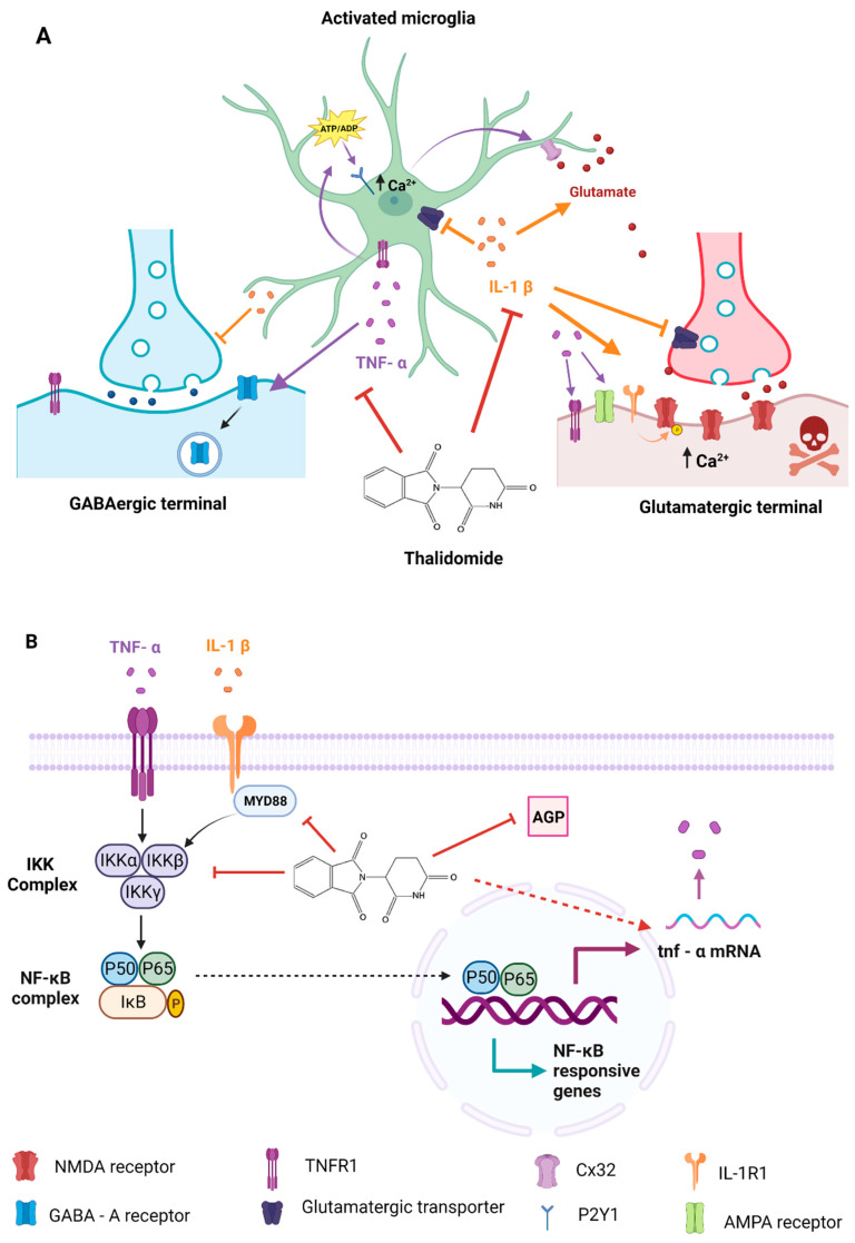
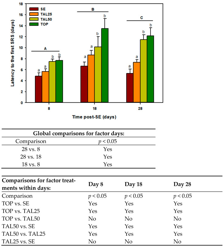
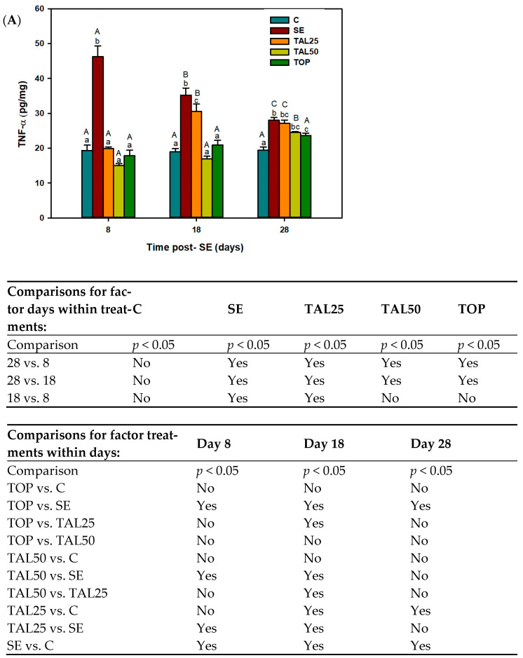
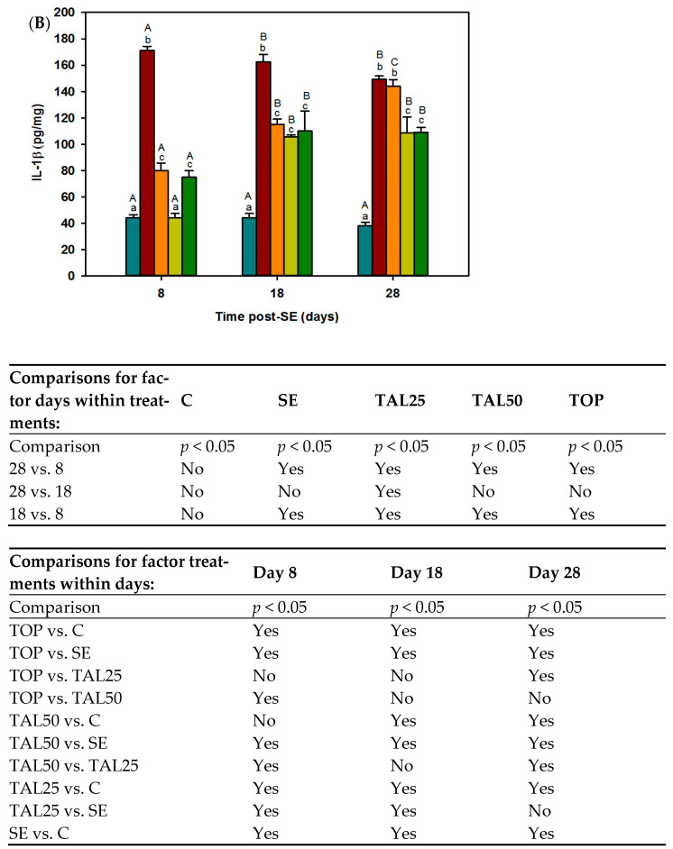
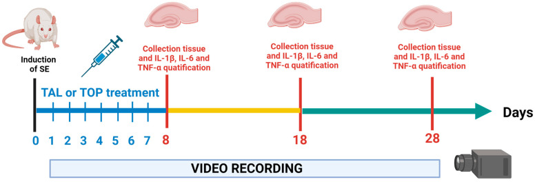
Thalidomide (TAL) has shown potential therapeutic effects in neurological diseases like epilepsy. Both clinical and preclinical studies show that TAL may act as an antiepileptic drug and as a possible treatment against disease development. However, the evidence for these effects is limited. Therefore, the antiepileptogenic and anti-inflammatory effects of TAL were evaluated herein. Sprague Dawley male rats were randomly allocated to one of five groups (n = 18 per group): control (C); status epilepticus (SE); SE-TAL (25 mg/kg); SE-TAL (50 mg/kg); and SE-topiramate (TOP; 60mg/kg). The lithium-pilocarpine model was used, and one day after SE induction the rats received pharmacological treatment for one week. The brain was obtained, and the hippocampus was micro-dissected 8, 18, and 28 days after SE. TNF-α, IL-6, and IL-1β concentrations were quantified. TOP and TAL (50 mg/kg) increased the latency to the first of many spontaneous recurrent seizures (SRS) and decreased SRS frequency, as well as decreasing TNF-α and IL-1β concentrations in the hippocampus. In conclusion, the results showed that both TAL (50 mg/kg) and TOP have anti-ictogenic and antiepileptogenic effects, possibly by decreasing neuroinflammation.

Keywords: anti-ictogenic; antiepileptogenic; neuroinflammation; temporal lobe epilepsy; thalidomide.


Conflict of interest statement

The authors declare no conflict of interest.

Figures

Figure 4
Figure 4
(A) Active microglia release proinflammatory cytokines, such as tumor necrosis factor alpha (TNF-α) and interleukin 1 beta (IL-1β). TNF-α induces microglial glutamate release through two mechanisms (1) from gap junction CX32 hemichannels [52] and (2) by activating the TNFR1 receptor in astroglia, which in turn release ATP/ADP to activate the P2Y1 purinergic receptor increasing the intracellular calcium concentration. Glutamate interacting with NMDA receptors promotes neuroexcitotoxicity [50,51]. TNF-α also induces GABAA receptor endocytosis and recruits AMPA receptors lacking GluR2 subunits, a conformation that favors the Ca2+ entry, amplifying the glutamate response [58] and activating TNFR1 neuronal receptors inducing cell death. IL-1β promotes NMDA receptor phosphorylation by interacting with its IL-1R1 receptor, which increases intracellular Ca2+ and excitotoxicity [75]. IL-1β can also increase glutamate concentrations by inhibiting its recapture by astrocytes and neurons [76,77], as well as by increasing astrocytic release [78,79,80]. Thalidomide (TAL) blocks IL-1β and TNF-α activity, decreasing neuronal damage and inflammation [34,35,73]. (B) Putative mechanism of action. TAL blocks TNF-α signaling by destabilizing the 3’unstranslated region (3´UTR) of TNF-α mRNA, inhibiting TNF-α protein synthesis [73]. TAL blocks nuclear factor kappa B (NF-κB) signaling which induces the expression of various pro-inflammatory genes by blocking the IKK complex [34,35] and via myeloid differentiating factor 88 (MyD88) [67]. Finally, TAL also can inhibit TNF-α by blocking α1-acid glycoprotein (AGP) [69]. Orange arrows represent the IL-1β processes while purple arrows represents the TNF-α processes. TAL mechanism are in red arrows. Imagen created with BioRender.com (accessed on 20 February 2023).
Figure 1
Figure 1
Effect of thalidomide (TAL) and topiramate (TOP) on the latency to the first spontaneous recurrent seizure observed 8, 18 and 28 days after status epilepticus (SE). Each bar represents the mean + S.E.M. of 6 animals per group. Different capital letters denote global comparisons p ≤ 0.05 among times, lowercase letters designate p ≤ 0.05 among treatments within times, two-way ANOVA followed by Student-Newman-Keuls post-hoc test. The tables show the results of statistical analysis.
Figure 2
Figure 2
Effect of thalidomide (TAL) and topiramate (TOP) on hippocampal proinflammatory cytokines quantified 8, 18 and 28 days after status epilepticus (SE). (A) TNF-α concentration of (B) IL-1β concentration and (C) IL-6 concentration. Each bar represents the mean + S.E.M. of 6 animals per group. Different capital letters denote p ≤ 0.05 among times within treatments, lowercase letters designate p ≤ 0.05 among treatments within times, two-way ANOVA followed by the Student-Newman-Keuls post-hoc test. C, control group. The tables show the results of statistical analysis.
Figure 2
Figure 2
Effect of thalidomide (TAL) and topiramate (TOP) on hippocampal proinflammatory cytokines quantified 8, 18 and 28 days after status epilepticus (SE). (A) TNF-α concentration of (B) IL-1β concentration and (C) IL-6 concentration. Each bar represents the mean + S.E.M. of 6 animals per group. Different capital letters denote p ≤ 0.05 among times within treatments, lowercase letters designate p ≤ 0.05 among treatments within times, two-way ANOVA followed by the Student-Newman-Keuls post-hoc test. C, control group. The tables show the results of statistical analysis.
Figure 3
Figure 3
(A) Correlation between TNF-α concentration and the latency to the first seizure in the TAL50 group at day 8 post-SE. (B) Correlation between TNF-α concentration and the number of SRS in the TOP group at 28 days post-SE. (C) Correlation between IL-1β concentration and the duration of SRS in the TAL25 group at 18 days post-SE. p < 0.05 Pearson correlation. (n = 6).
Figure 3
Figure 3
(A) Correlation between TNF-α concentration and the latency to the first seizure in the TAL50 group at day 8 post-SE. (B) Correlation between TNF-α concentration and the number of SRS in the TOP group at 28 days post-SE. (C) Correlation between IL-1β concentration and the duration of SRS in the TAL25 group at 18 days post-SE. p < 0.05 Pearson correlation. (n = 6).
Figure 5
Figure 5
Experimental design. At time 0, status epilepticus (SE) was induced in male Sprague-Dawley rats via administration of lithium-pilocarpine. One day after SE induction rats received daily pharmacological treatment with topiramate (TOP) or thalidomide (TAL) for seven days. One day after treatment the first determination was performed (evaluation time 8). The animals were sacrificed, and hippocampus were collected to determine the concentrations of interleukin IL-1β, IL-6 and necrosis tumoral factor-α (TNF-α) at three different evaluation times: 8, 18 and 28 post-SE days respectively. From 1 to 28 days post-SE, rats were video monitored during 24 h/7 days to observe the latency to the first spontaneous recurrent seizure (SRS) as well as, SRS number and duration.

Similar articles

Cited by

References

    1. Engel J., Jr. Mesial Temporal Lobe Epilepsy: What Have We Learned? Neuroscientist. 2001;7:340–352. doi: 10.1177/107385840100700410. - DOI - PubMed
    1. Englot D.J., Morgan V.L., Chang C. Impaired vigilance networks in temporal lobe epilepsy: Mechanisms and clinical implications. Epilepsia. 2020;61:189–202. doi: 10.1111/epi.16423. - DOI - PMC - PubMed
    1. Curia G., Longo D., Biagini G., Jones R.S., Avoli M. The pilocarpine model of temporal lobe epilepsy. J. Neurosci. Methods. 2008;172:143–157. doi: 10.1016/j.jneumeth.2008.04.019. - DOI - PMC - PubMed
    1. Tatum W.O. Mesial Temporal Lobe Epilepsy. J. Clin. Neurophysiol. 2012;29:356–365. doi: 10.1097/WNP.0b013e31826b3ab7. - DOI - PubMed
    1. Scorza F., Arida R.M., Mazzacoratti M.D.G.N., Scerni D.A., Calderazzo L., Cavalheiro E.A. The pilocarpine model of epilepsy: What have we learned? An. Acad. Bras. Cienc. 2009;81:345–365. doi: 10.1590/S0001-37652009000300003. - DOI - PubMed

MeSH terms