. 2023 Mar 31;24(7):6536.
doi: 10.3390/ijms24076536.

Effects of Follicle-Stimulating Hormone on Human Sperm Motility In Vitro

Affiliations

Effects of Follicle-Stimulating Hormone on Human Sperm Motility In Vitro

Rossella Cannarella et al. Int J Mol Sci. .

Abstract

To evaluate whether the follicle-stimulating hormone (FSH) receptor (FSHR) is expressed in human spermatozoa and the effects of FSH incubation on sperm function. Twenty-four Caucasian men were recruited. Thirteen patients had asthenozoospermia, and the remaining 11 had normal sperm parameters (controls). After confirming FSHR expression, spermatozoa from patients and controls were incubated with increasing concentrations of human purified FSH (hpFSH) to reassess FSHR expression and localization and to evaluate progressive and total sperm motility, the mitochondrial membrane potential, and protein kinase B (AKT) 473 and 308 phosphorylation. FSHR is expressed in the post-acrosomal segment, neck, midpiece, and tail of human spermatozoa. Its localization does not differ between patients and controls. Incubation with hpFSH at a concentration of 30 mIU/mL appeared to increase FSHR expression mainly in patients. Incubation of human spermatozoa with hpFSH overall resulted in an overall deterioration of both progressive and total motility in patients and controls and worse mitochondrial function only in controls. Finally, incubation with FSH increased AKT473/tubulin phosphorylation to a greater extent than AKT308. FSHR is expressed in the post-acrosomal region, neck, midpiece, and tail of human spermatozoa. Contrary to a previous study, we report a negative effect of FSH on sperm motility and mitochondrial function. FSH also activates the AKT473 signaling pathway.

Keywords: FSH; FSHR; sperm motility; spermatozoa.


Conflict of interest statement

The authors declare no conflict of interest.

Figures

Figure 1
Figure 1
Immunolocalization of the follicle-stimulating hormone receptor (FSHR) protein in human spermatozoa. Immunofluorescence (IF) analysis of spermatozoa. The protein is present in the post-acrosomal segment, neck, midpiece, and tail of the spermatozoon. Magnification 100×. IF is indicated by the red arrow.
Figure 2
Figure 2
Expression of follicle-stimulating hormone receptor (FSHR) protein in human spermatozoa from patients and controls before and after incubation with hpFSH. Western blot (WB) analysis of FSHR in positive [Sertoli cells (SCs)] and negative (HT29) controls (A). WB analysis of FSHR in spermatozoa from patients and controls (B). WB analysis of tubulin in spermatozoa from patients and controls (C). Densitometric analysis of the FSHR/tubulin ratio in spermatozoa from controls (D). Densitometric analysis of the FSHR/tubulin ratio in spermatozoa from patients (E). The data represent the mean ± standard deviation of three independent experiments, each performed in triplicate.
Figure 3
Figure 3
Immunolocalization of the follicle-stimulating hormone receptor (FSHR) protein in human spermatozoa after incubation with hpFSH. Immunofluorescence (IF) analysis of the FSHR in spermatozoa of controls and patients (A). Magnification from Patient 4: before incubation, the protein was not identified (Left panel). After incubation, it appeared in the post-acrosomal region, neck, midpiece, and tail (Right panel) (B). Magnification from Control 2 (Left panel) and Patient 2 (Right panel): the protein appears expressed even before incubation with FSH (C). Magnification 100×. IF is indicated by the red arrows in Panels B and C.
Figure 3
Figure 3
Immunolocalization of the follicle-stimulating hormone receptor (FSHR) protein in human spermatozoa after incubation with hpFSH. Immunofluorescence (IF) analysis of the FSHR in spermatozoa of controls and patients (A). Magnification from Patient 4: before incubation, the protein was not identified (Left panel). After incubation, it appeared in the post-acrosomal region, neck, midpiece, and tail (Right panel) (B). Magnification from Control 2 (Left panel) and Patient 2 (Right panel): the protein appears expressed even before incubation with FSH (C). Magnification 100×. IF is indicated by the red arrows in Panels B and C.
Figure 4
Figure 4
Effects of increasing concentrations of follicle-stimulating hormone (FSH) on in-toto semen sample (A), semen pellet (B), and spermatozoa recovered by swim-up (C) from normozoospermic men and asthenozoospermic patients. Data are reported as the percentage change from the control (FSH 0 mIU/mL), using boxplots. The line inside the box indicates the median, the end of the box the interquartile range, and the values outside the box show the minimum and maximum values. The different colors indicate the different doses of FSH used for sample incubation. * p < 0.05 vs. 0 mIU/mL;  p < 0.05 vs. 1 mIU/mL.
Figure 5
Figure 5
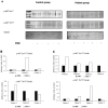
Phosphorylation of protein kinase B (AKT) in Serine 473 and Threonine 308. Immunoblots (A) and densitometric analysis (B,C) of p-AKT Ser473/tubulin ratio and p-AKT Thr308/tubulin ratio in patients and controls. p-AKT Ser473/Tubulin increased after incubation with hpFSH in most of the patients and controls. p-AKT Ser308/tubulin ratio increased in a minority of patients and controls.
Figure 6
Figure 6
Flow chart of the experimental design. After confirmation of follicle-stimulating hormone receptor (FSHR) expression in human spermatozoa by polymerase chain reaction (PCR), Western blot, and immunofluorescence, experiments were conducted in men with normozoospermia and patients with asthenozoospermia using spermatozoa separated by swim-up, in-toto semen, or semen pellets.

Similar articles

Cited by

References

    1. Khanehzad M., Abbaszadeh R., Holakuyee M., Modarressi M.H., Nourashrafeddin S.M. FSH regulates RA signaling to commit spermatogonia into differentiation pathway and meiosis. Reprod. Biol. Endocrinol. 2021;19:4. doi: 10.1186/s12958-020-00686-w. - DOI - PMC - PubMed
    1. Hofmann M.C. Gdnf signaling pathways within the mammalian spermatogonial stem cell niche. Mol. Cell. Endocrinol. 2008;288:95–103. doi: 10.1016/j.mce.2008.04.012. - DOI - PMC - PubMed
    1. Johnston D.S., Olivas E., Di Candeloro P., Wright W.W. Stage-specific changes in GDNF expression by rat Sertoli cells: A possible regulator of the replication and differentiation of stem spermatogonia. Biol. Reprod. 2011;85:763–769. doi: 10.1095/biolreprod.110.087676. - DOI - PMC - PubMed
    1. Quintana J., Hipkin R.W., Ascoli M. A polyclonal antibody to a synthetic peptide derived from the rat follicle-stimulating hormone receptor reveals the recombinant receptor as a 74-kilodalton protein. Endocrinology. 1993;133:2098–2104. doi: 10.1210/endo.133.5.8404659. - DOI - PubMed
    1. Davis D., Liu X., Segaloff D.L. Identification of the sites of N-linked glycosylation on the follicle-stimulating hormone (FSH) receptor and assessment of their role in FSH receptor function. Mol. Endocrinol. 1995;9:159–170. - PubMed

Substances

LinkOut - more resources