. 2023 Mar 31;24(7):6550.
doi: 10.3390/ijms24076550.

Nyamanini Virus Nucleoprotein and Phosphoprotein Organize Viral Inclusion Bodies That Associate with Host Biomolecular Condensates in the Nucleus

Affiliations

Nyamanini Virus Nucleoprotein and Phosphoprotein Organize Viral Inclusion Bodies That Associate with Host Biomolecular Condensates in the Nucleus

Yuya Hirai et al. Int J Mol Sci. .

Abstract

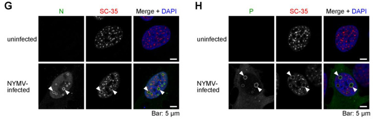
Many mononegaviruses form inclusion bodies (IBs) in infected cells. However, little is known about nuclear IBs formed by mononegaviruses, since only a few lineages of animal-derived mononegaviruses replicate in the nucleus. In this study, we characterized the IBs formed by Nyamanini virus (NYMV), a unique tick-borne mononegavirus undergoing replication in the nucleus. We discovered that NYMV forms IBs, consisting of condensates and puncta of various sizes and morphologies, in the host nucleus. Likewise, we found that the expressions of NYMV nucleoprotein (N) and phosphoprotein (P) alone induce the formation of condensates and puncta in the nucleus, respectively, even though their morphologies are somewhat different from the IBs observed in the actual NYMV-infected cells. In addition, IB-like structures can be reconstructed by co-expressions of NYMV N and P, and localization analyses using a series of truncated mutants of P revealed that the C-terminal 27 amino acid residues of P are important for recruiting P to the condensates formed by N. Furthermore, we found that nuclear speckles, cellular biomolecular condensates, are reorganized and recruited to the IB-like structures formed by the co-expressions of N and P, as well as IBs formed in NYMV-infected cells. These features are unique among mononegaviruses, and our study has contributed to elucidating the replication mechanisms of nuclear-replicating mononegaviruses and the virus-host interactions.

Keywords: Nyamanini virus; biomolecular condensates; liquid–liquid phase separation; membraneless organelles; mononegavirus; non-segmented negative-strand RNA viruses; nuclear speckles; nucleoprotein; nucleus; phosphoprotein.


Conflict of interest statement

The authors declare no conflict of interest.

Figures

Figure 1
Figure 1
The formation of IBs-like structures by NMYV N and P. (A) A schematic diagram of the genome structure of NYMV. The gene regions of N and P are highlighted in grey. (B,C) The localizations of N (B) and P (C) in NYMV-infected U-2 OS cells revealed by confocal immunofluorescence microscopy using specific antibodies. (D) The localization of N or P that was exogenously expressed by transfection (TF) in uninfected U-2 OS cells revealed by confocal immunofluorescence microscopy using anti-N or anti-P antibodies, respectively. (E) The localizations of N in uninfected U-2 OS cells that were transfected with N and FLAG-tag (upper) or N and FLAG-tagged P (lower). Arrowheads and arrows indicate condensates and puncta, respectively. (F) The number of condensates in the nucleus in (E). n > 300 from three samples. Statistical significance was assessed by using a two-tailed Welch’s t-test. The confocal microscopic images presented in (BE) were the images of single focal planes.
Figure 2
Figure 2
Subcellular localizations of a series of deletion mutants of NYMV P. (A) A schematic diagram of a series of deletion mutants of P used in this experiment and the summary of the localizations of P mutants and N when co-expressed with P mutants. Numbers 1–382 represents the full-length P. (B) The localizations of a series of deletion mutants of P in uninfected U-2 OS cells in the case of P mutants alone (left column) or when co-expressed with N (right column). Arrowheads and arrows indicate condensates and puncta, respectively. The presented confocal microscopic images were the images of single focal planes.
Figure 3
Figure 3
Minireplicon assay using truncated NYMV P. Minireplicon assay was performed on P mutants. The relative luciferase activities are indicated (biological replicates; n = 3). The line indicates the average, and blue dots represent individual data. NC, negative control; 1–382, full-length P; 86–382, P86–382; 1–355, P1–355.
Figure 4
Figure 4
The relationship between viral IB-like structures of NYMV and the nuclear biomolecular condensates. (AD) N alone, P alone, or both N and P were expressed in uninfected U-2 OS cells, and their co-localizations with nucleoli recognized by an anti-B23 antibody (A,B) or nuclear speckles recognized by an anti-SC-35 antibody (C,D) were examined. Arrowheads indicate abnormal nucleoli co-localized with viral condensates in (A,B). Arrowheads and arrows indicate nuclear speckle co-localized with viral condensates and puncta, respectively in (C,D). (EH) The co-localizations of N and P with nucleoli (E,F) or nuclear speckles (G,H) in NYMV-infected U-2 OS cells were examined. Arrowheads indicate nuclear speckles co-localized with IBs in (G,H). The presented confocal microscopic images were the images of single focal planes.
Figure 4
Figure 4
The relationship between viral IB-like structures of NYMV and the nuclear biomolecular condensates. (AD) N alone, P alone, or both N and P were expressed in uninfected U-2 OS cells, and their co-localizations with nucleoli recognized by an anti-B23 antibody (A,B) or nuclear speckles recognized by an anti-SC-35 antibody (C,D) were examined. Arrowheads indicate abnormal nucleoli co-localized with viral condensates in (A,B). Arrowheads and arrows indicate nuclear speckle co-localized with viral condensates and puncta, respectively in (C,D). (EH) The co-localizations of N and P with nucleoli (E,F) or nuclear speckles (G,H) in NYMV-infected U-2 OS cells were examined. Arrowheads indicate nuclear speckles co-localized with IBs in (G,H). The presented confocal microscopic images were the images of single focal planes.

Similar articles

References

    1. Cifuentes-Muñoz N., Branttie J., Slaughter K.B., Dutch R.E. Human Metapneumovirus Induces Formation of Inclusion Bodies for Efficient Genome Replication and Transcription. J. Virol. 2017;91:e01282-17. doi: 10.1128/JVI.01282-17. - DOI - PMC - PubMed
    1. Lahaye X., Vidy A., Pomier C., Obiang L., Harper F., Gaudin Y., Blondel D. Functional Characterization of Negri Bodies (NBs) in Rabies Virus-Infected Cells: Evidence That NBs Are Sites of Viral Transcription and Replication. J. Virol. 2009;83:7948–7958. doi: 10.1128/JVI.00554-09. - DOI - PMC - PubMed
    1. Heinrich B.S., Cureton D.K., Rahmeh A.A., Whelan S.P.J. Protein Expression Redirects Vesicular Stomatitis Virus RNA Synthesis to Cytoplasmic Inclusions. PLoS Pathog. 2010;6:e1000958. doi: 10.1371/journal.ppat.1000958. - DOI - PMC - PubMed
    1. Hoenen T., Shabman R.S., Groseth A., Herwig A., Weber M., Schudt G., Dolnik O., Basler C.F., Becker S., Feldmann H. Inclusion Bodies Are a Site of Ebolavirus Replication. J. Virol. 2012;86:11779–11788. doi: 10.1128/JVI.01525-12. - DOI - PMC - PubMed
    1. Ringel M., Heiner A., Behner L., Halwe S., Sauerhering L., Becker N., Dietzel E., Sawatsky B., Kolesnikova L., Maisner A. Nipah Virus Induces Two Inclusion Body Populations: Identification of Novel Inclusions at the Plasma Membrane. PLoS Pathog. 2019;15:e1007733. doi: 10.1371/journal.ppat.1007733. - DOI - PMC - PubMed

MeSH terms

LinkOut - more resources