

Three-Dimensional Bioprinting of Organoid-Based Scaffolds (OBST) for Long-Term Nanoparticle Toxicology Investigation
- PMID: 37047568
- PMCID: PMC10095512
- DOI: 10.3390/ijms24076595
Three-Dimensional Bioprinting of Organoid-Based Scaffolds (OBST) for Long-Term Nanoparticle Toxicology Investigation
Abstract
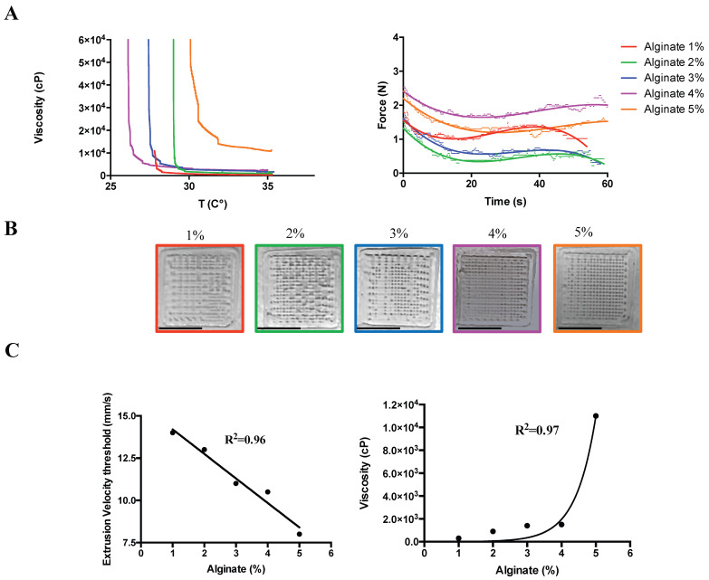
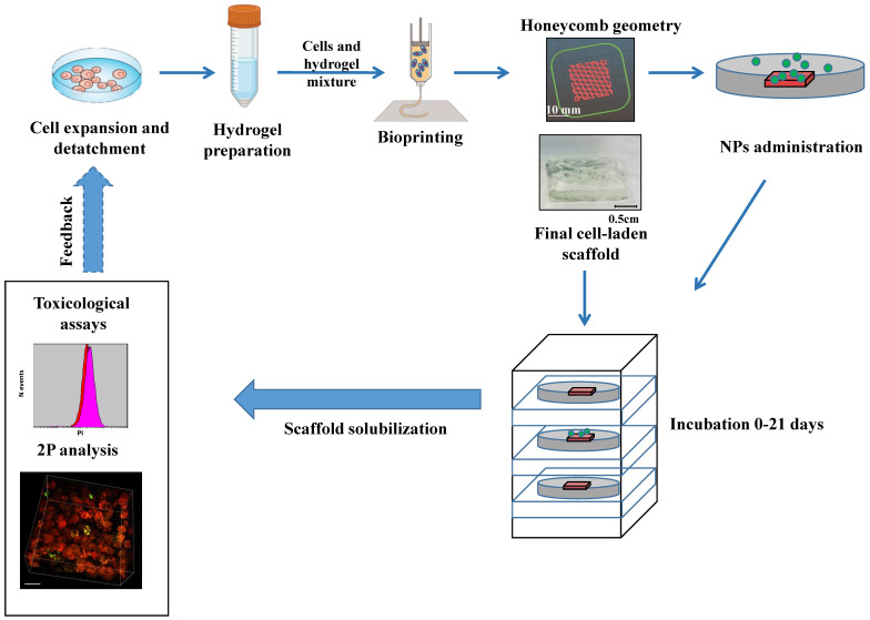
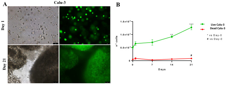
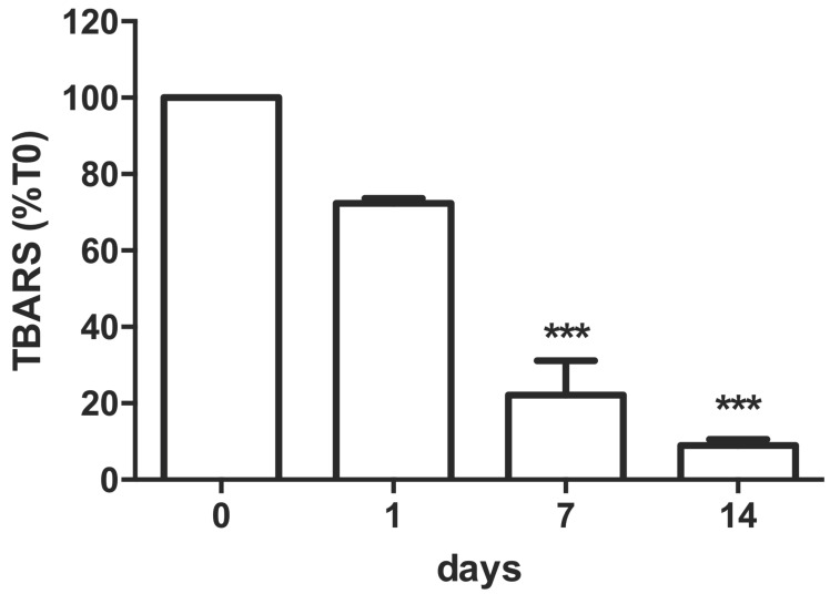
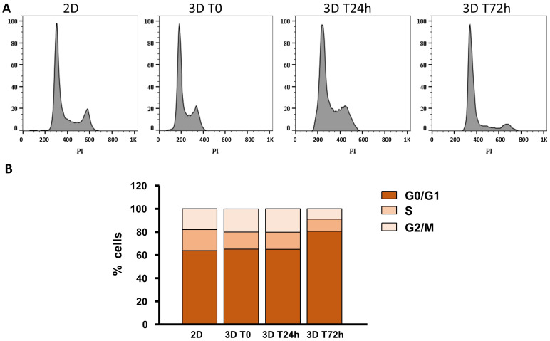
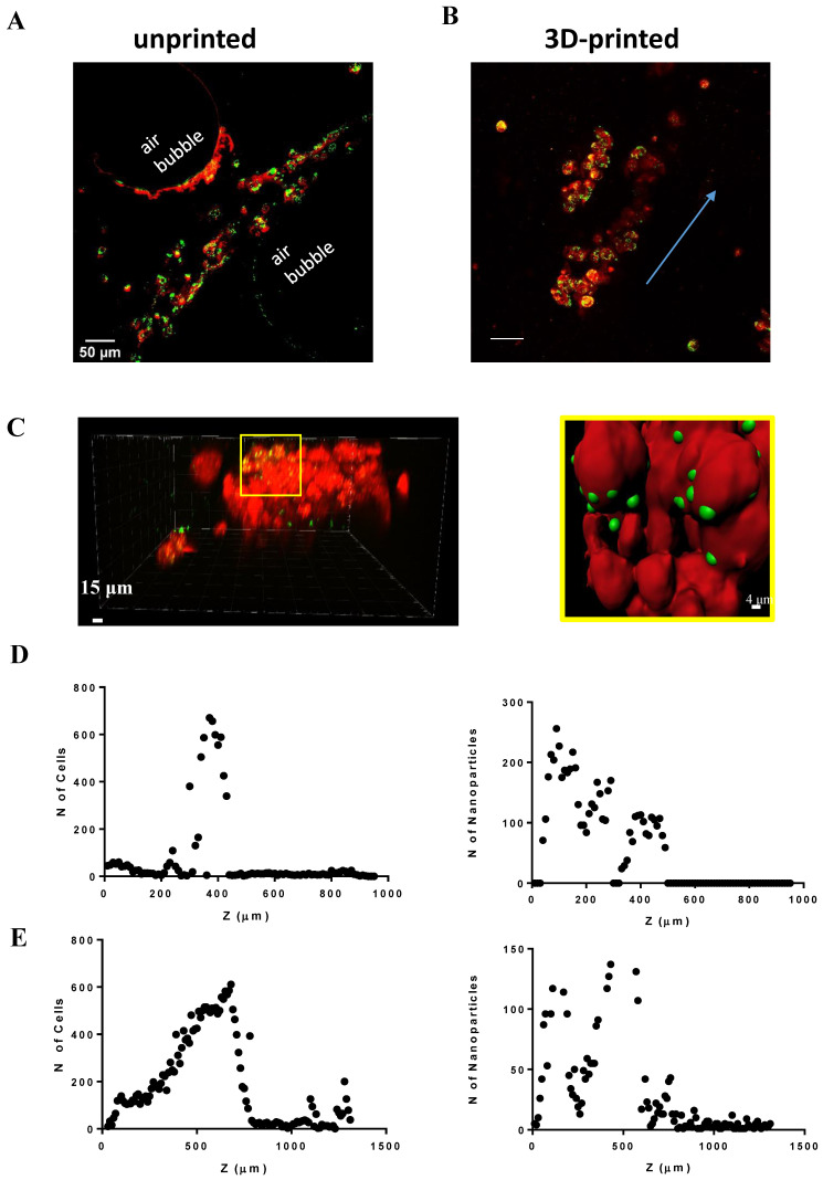
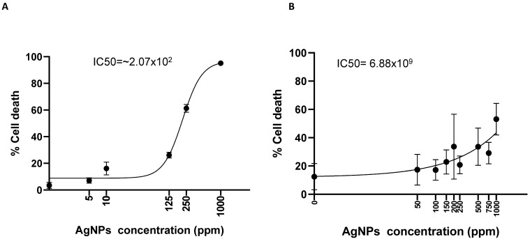
The toxicity of nanoparticles absorbed through contact or inhalation is one of the major concerns for public health. It is mandatory to continually evaluate the toxicity of nanomaterials. In vitro nanotoxicological studies are conventionally limited by the two dimensions. Although 3D bioprinting has been recently adopted for three-dimensional culture in the context of drug release and tissue regeneration, little is known regarding its use for nanotoxicology investigation. Therefore, aiming to simulate the exposure of lung cells to nanoparticles, we developed organoid-based scaffolds for long-term studies in immortalized cell lines. We printed the viscous cell-laden material via a customized 3D bioprinter and subsequently exposed the scaffold to either 40 nm latex-fluorescent or 11-14 nm silver nanoparticles. The number of cells significantly increased on the 14th day in the 3D environment, from 5 × 10⁵ to 1.27 × 10⁶, showing a 91% lipid peroxidation reduction over time and minimal cell death observed throughout 21 days. Administered fluorescent nanoparticles can diffuse throughout the 3D-printed scaffolds while this was not the case for the unprinted ones. A significant increment in cell viability from 3D vs. 2D cultures exposed to silver nanoparticles has been demonstrated. This shows toxicology responses that recapitulate in vivo experiments, such as inhaled silver nanoparticles. The results open a new perspective in 3D protocols for nanotoxicology investigation supporting 3Rs.
Keywords: 3D bioprinter; Calu-3; long-term culture; nanoparticles; nanotoxicology.
Conflict of interest statement
Ruben Foresti is the founder and shareholder of UIMEI Srl, a company that develops 3D printers and medical devices. Nevertheless, he does not gain or lose financially through publication. The authors have no potential conflict of interest.
Figures
Similar articles
-
Three-Dimensional Bioprinting of Organoids: Past, Present, and Prospective.Tissue Eng Part A. 2024 Jun;30(11-12):314-321. doi: 10.1089/ten.TEA.2023.0209. Epub 2024 Feb 2. Tissue Eng Part A. 2024. PMID: 38205663 Review.
-
3D-bioprinting of aortic valve interstitial cells: impact of hydrogel and printing parameters on cell viability.Biomed Mater. 2022 Nov 11;18(1). doi: 10.1088/1748-605X/ac9f91. Biomed Mater. 2022. PMID: 36322974
-
Alginate dependent changes of physical properties in 3D bioprinted cell-laden porous scaffolds affect cell viability and cell morphology.Biomed Mater. 2019 Sep 25;14(6):065009. doi: 10.1088/1748-605X/ab3c74. Biomed Mater. 2019. PMID: 31426033
-
Advanced roll porous scaffold 3D bioprinting technology.J Artif Organs. 2025 Jun;28(2):225-233. doi: 10.1007/s10047-024-01470-y. Epub 2024 Sep 27. J Artif Organs. 2025. PMID: 39327399 Review.
-
3D Biofabrication of a Cardiac Tissue Construct for Sustained Longevity and Function.ACS Appl Mater Interfaces. 2022 May 18;14(19):21800-21813. doi: 10.1021/acsami.1c23883. Epub 2022 May 9. ACS Appl Mater Interfaces. 2022. PMID: 35533308 Free PMC article.
Cited by
-
(3D) Bioprinting-Next Dimension of the Pharmaceutical Sector.Pharmaceuticals (Basel). 2024 Jun 17;17(6):797. doi: 10.3390/ph17060797. Pharmaceuticals (Basel). 2024. PMID: 38931464 Free PMC article. Review.
-
Surgical Medical Education via 3D Bioprinting: Modular System for Endovascular Training.Bioengineering (Basel). 2024 Feb 19;11(2):197. doi: 10.3390/bioengineering11020197. Bioengineering (Basel). 2024. PMID: 38391683 Free PMC article.
-
The Upper Limb Orthosis in the Rehabilitation of Stroke Patients: The Role of 3D Printing.Bioengineering (Basel). 2023 Oct 27;10(11):1256. doi: 10.3390/bioengineering10111256. Bioengineering (Basel). 2023. PMID: 38002380 Free PMC article. Review.
-
Bioprinting of Cells, Organoids and Organs-on-a-Chip Together with Hydrogels Improves Structural and Mechanical Cues.Cells. 2024 Oct 1;13(19):1638. doi: 10.3390/cells13191638. Cells. 2024. PMID: 39404401 Free PMC article. Review.
-
A Futuristic Development in 3D Printing Technique Using Nanomaterials with a Step Toward 4D Printing.ACS Omega. 2024 Aug 26;9(36):37445-37458. doi: 10.1021/acsomega.4c04123. eCollection 2024 Sep 10. ACS Omega. 2024. PMID: 39281933 Free PMC article. Review.
References
-
- Marrella A., Iafisco M., Adamiano A., Rossi S., Aiello M., Barandalla-Sobrados M., Carullo P., Miragoli M., Tampieri A., Scaglione S., et al. A combined low-frequency electromagnetic and fluidic stimulation for a controlled drug release from superparamagnetic calcium phosphate nanoparticles: Potential application for cardiovascular diseases. J. R. Soc. Interface. 2018;15:20180236. doi: 10.1098/rsif.2018.0236. - DOI - PMC - PubMed
MeSH terms
Substances
LinkOut - more resources
Full Text Sources
