. 2023 Apr 2;24(7):6639.
doi: 10.3390/ijms24076639.

The Labyrinthine Landscape of APP Processing: State of the Art and Possible Novel Soluble APP-Related Molecular Players in Traumatic Brain Injury and Neurodegeneration

Affiliations

The Labyrinthine Landscape of APP Processing: State of the Art and Possible Novel Soluble APP-Related Molecular Players in Traumatic Brain Injury and Neurodegeneration

Mirco Masi et al. Int J Mol Sci. .

Abstract

Amyloid Precursor Protein (APP) and its cleavage processes have been widely investigated in the past, in particular in the context of Alzheimer's Disease (AD). Evidence of an increased expression of APP and its amyloidogenic-related cleavage enzymes, β-secretase 1 (BACE1) and γ-secretase, at the hit axon terminals following Traumatic Brain Injury (TBI), firstly suggested a correlation between TBI and AD. Indeed, mild and severe TBI have been recognised as influential risk factors for different neurodegenerative diseases, including AD. In the present work, we describe the state of the art of APP proteolytic processing, underlining the different roles of its cleavage fragments in both physiological and pathological contexts. Considering the neuroprotective role of the soluble APP alpha (sAPPα) fragment, we hypothesised that sAPPα could modulate the expression of genes of interest for AD and TBI. Hence, we present preliminary experiments addressing sAPPα-mediated regulation of BACE1, Isthmin 2 (ISM2), Tetraspanin-3 (TSPAN3) and the Vascular Endothelial Growth Factor (VEGFA), each discussed from a biological and pharmacological point of view in AD and TBI. We finally propose a neuroprotective interaction network, in which the Receptor for Activated C Kinase 1 (RACK1) and the signalling cascade of PKCβII/nELAV/VEGF play hub roles, suggesting that vasculogenic-targeting therapies could be a feasible approach for vascular-related brain injuries typical of AD and TBI.

Keywords: BACE1; ELAV; ISM2; PKC; RACK1; TSPAN3; VEGF; secretase.


Conflict of interest statement

The authors declare no conflict of interests.

Figures

Figure 1
Figure 1
APP gene, mRNA and protein structure. (a) Structure of APP gene and mRNA. APP gene, located on chromosome 21q21.3, features 18 exons. Alternative splicing of exons 7 and 8 (dark grey) leads to the expression of APP695, 751 and 770 major isoforms, while differential splicing of exons 2 and 15 (light grey) generates APP639 and L-APP, respectively. (b) Structure of the three APP protein family members APP, APLP1 and APLP2. From the N-terminus to the C-terminus, APP features the cysteine-rich E1 domain (with Heparin-Binding Site (HBS), a Zinc-Binding Site (ZnBS) and Copper-Binding Site (CuBS)), the Extension Domain (ED), the Acidic Domain (AcD), the helix-rich E2 domain (with a second HBS and a Collagen-Binding Site (CBS)), the Juxtamembrane Region (JMR), the Aβ sequence, the Transmembrane Domain (TM) and APP Intracellular Domain (AICD) which contains a YENPTY sorting motif. APP751 and APP770 contain the additional Kunitz Protease Inhibitor (KPI) domain and an OX-2 antigen domain. Amino-acid sequence of Aβ region is shown along with the different secretases cleavage sites as well as the Aβ product lines. Both APLP1 and APLP2 lack Aβ sequence and present the APLP-intracellular domain 1 (ALID1) and ALID2, respectively. APLP2 features a KPI domain similarly to some APP isoforms.
Figure 2
Figure 2
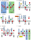
Schematic overview of APP-processing pathways. (a) APP canonical proteolytic processing. In the canonical cleavage, APP is either processed in the non-amyloidogenic pathway, where the sequential cleavage by α-secretase (ADAM10) (blue square) and the γ-secretase complex (purple square) produces sAPPα and p3, or in the amyloidogenic pathway, where the sequential cleavage by β-secretase 1 (green square) or Cathepsin B and the γ-secretase complex liberates sAPPβ and Aβ. While α-produced AICD are rapidly degraded in the cytoplasm, β-generated AICD form the transcriptional factor ATF complex together with Fe65 and Tip60, that translocates into the nucleus to up-regulate APP-related genes (see text for details). (bf) APP non-canonical proteolytic processing. (b) APP processing mediated by δ-secretase (orange square) releases three soluble APP fragments (sAPP1–585, sAPP1–373, and sAPP374–585) and δ-CTF, which is further processed by β- and γ-secretases, releasing Aβ, AICD and the C586–695 fragment. (c) η-secretase (light green square) releases sAPPη and η-CTF, which is further processed by either α- or β-secretase 1, releasing Aη-α or Aη-β respectively; the remaining CTFs are cleaved by the γ-secretase complex, releasing p3 and AICD, or Aβ and AICD respectively. (d) Meprin-β (brown rectangle) produces sAPPβ* (similar to sAPPβ) and two shorter soluble fragments (sAPP1–124 and sAPP1–380/3), while the remaining β*-CTF is processed by the γ-secretase complex, releasing AICD and Aβ2–X. (e) θ-secretase (grey square) can either cleave APP at the θ-site, releasing sAPPθ and a θ-CTF that is further processed by the γ-secretase complex releasing AICD and a truncated form of Aβ, or act as a conditional β-secretase. (f) Caspase-3, -6 and -8 (yellow rectangle) cleave within APP Intracellular Domain, releasing C31 while, after the sequential cleavage operated by the γ-secretase complex, the small peptide JCasp is released in the cytoplasm.
Figure 3
Figure 3
Alteration of APP processing after TBI. (a) Acute axonal damage after TBI induces the expression of APP, β-secretase 1 and γ-secretase components PS1 and PS2 (dark green arrow), as well as an increased Aβ generation and deposition. In addition, TBI triggers C/EBPβ activation, which induces the expression of δ-secretase (dark green arrow) that, in turn, cleaves APP forming Aβ and the C586–695 peptide that activates C/EBPβ. This suggests the establishment of a deleterious vicious loop possibly correlated to AD development. (b) Epigenetic and mechanical components of TBI-triggered APP processing alterations. After TBI, a differential CpG methylation of APP, MAPT, Neurofilament Heavy (NEFH), Neurofilament Medium (NEFM) and Neurofilament Light (NEFL) is observed [92]. Moreover, the induced axonal cytoskeleton distortion impairs the fast-axonal transport of APP (light green arrow), which accumulates in proximity of the axonal injury site, resulting in Aβ deposition.
Figure 4
Figure 4
Effects of sAPPα treatment on early MAPK activation and BACE1, TSPAN3 and ISM2 transcriptional regulation. Human neuroblastoma SH-SY5Y cells were cultured and treated with 10 nM sAPPα for 15, 30 or 60 min (ac) or for 24 h (df) as previously detailed [141]. Western blot analysis and qPCR were performed as previously reported [141]. (ac) Evaluation of early MAPK activation. The image is a representative Western blot. Phosphorylation of p42 and p44 was normalised to their respective total p42 and p44 levels. (df) Evaluation of sAPPα-mediated transcriptional regulation on BACE1, TSPAN3 and ISM2. mRNA levels were evaluated by qPCR (endogenous reference, RPL6). Results are expressed as mean ± SEM, n = 3 independent experiments. Statistical analysis was performed with one-way ANOVA followed by Dunnett’s multiple comparison test (b,c) or with Student’s t-test (df) with * p < 0.05 and ** p < 0.01 vs. control (CTRL). Statistical significance is detailed in the respective figure panel.
Figure 5
Figure 5
Effects of sAPPα treatment on VEGF expression. SH-SY5Y cells were treated with 10 nM sAPPα for 6 or 24 h. (a) SH-SY5Y cells transiently transfected with Δ1_VEGF luciferase-reporter plasmid and treated with sAPPα were lysed and luciferase activity was measured as previously described [141]. Luciferase activity is expressed as RLU% normalised to non-treated construct (set as 100%). (b,c) Evaluation of sAPPα-mediated VEGFA transcriptional regulation. mRNA levels were assessed by qPCR and normalised on RPL6 (b) or the respective VEGFA isoform (employed primers described in [206]) (c). (d) Evaluation of sAPPα effects on VEGFA mRNA stability. SH-SY5Y cells were pre-treated with 50 µM DRB 53-85-0 (a classic RNA polymerase II inhibitor), then treated with 10 nM sAPPα for 0, 4, 6 or 8 h. RNA was extracted and reverse-transcribed as previously described [141]. mRNA levels were assessed by qPCR. Results are expressed as a percentage of the initial steady-state VEGFA mRNA levels. (e) Evaluation of VEGF protein levels in cell lysates and supernatants (SN). The image is a representative Western blot. Results are expressed as mean ± SEM, n = 3 independent experiments. Statistical analysis was performed with one-way ANOVA followed by Dunnett’s multiple comparison test (ac), with Bonferroni multiple comparison test (d) or Student’s t-test (e) with * p < 0.05 and ** p < 0.01 vs. control (CTRL), *** p < 0.001 DRB vs. DRB + sAPPα (6 h) and §§ p < 0.01 DRB vs. DRB + sAPPα (8 h). Statistical significance is detailed in the respective figure panel.
Figure 6
Figure 6
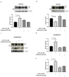
Evaluation of the PKCβII/nELAV/VEGF pathway in sAPPα-mediated effects on VEGF. (ae) SH-SY5Y cells were treated with 10 nM sAPPα alone or in combination with 0.2 µM Wortmannin (an irreversible PI3K inhibitor). Subcellular fractionation was performed as previously described [141]. The image is a representative Western blot. nELAV protein levels were analysed in the nucleus (a), cytosol (b) and cytoskeleton (c,d) fractions. PKCβII protein levels were analysed in the cytoskeleton fraction (c,e). Protein levels were normalised to α-tubulin expression. Results are expressed as mean ± SEM, n = 3 independent experiments. Statistical analysis was performed with one-way ANOVA followed by Dunnett’s multiple comparison test with * p < 0.05 and ** p < 0.01 vs. control (untreated cells). Statistical significance is detailed in the respective figure panel.
Figure 7
Figure 7
Putative sAPPα-related VEGF-RACK1 neuroprotective interaction network. RACK1 has been demonstrated to play pivotal roles in neuronal context and its modulation has been observed to be influenced by sAPPα treatment. Therefore, considering the reported and putative interactions among RACK1 and several players here discussed, it is possible to hypothesise a molecular network correlated to sAPPα and with potential implications in both AD and TBI context, from both a biological and pharmacological point of view (bold lines = protein-protein interactions or protein functional correlations reported in literature and cited in the text; dash lines = putative structural and/or functional protein correlations hypothesised based on available literature data).

Similar articles

Cited by

References

    1. Wolozin B., Ivanov P. Stress granules and neurodegeneration. Nat. Rev. Neurosci. 2019;20:649–666. doi: 10.1038/s41583-019-0222-5. - DOI - PMC - PubMed
    1. Masi M., Attanzio A., Racchi M., Wolozin B., Borella S., Biundo F., Buoso E. Proteostasis Deregulation in Neurodegeneration and Its Link with Stress Granules: Focus on the Scaffold and Ribosomal Protein RACK1. Cells. 2022;11:2590. doi: 10.3390/cells11162590. - DOI - PMC - PubMed
    1. Hardy J., Selkoe D.J. The amyloid hypothesis of Alzheimer’s disease: Progress and problems on the road to therapeutics. Science. 2002;297:353–356. doi: 10.1126/science.1072994. - DOI - PubMed
    1. Zheng H., Koo E.H. Biology and pathophysiology of the amyloid precursor protein. Mol. Neurodegener. 2011;6:27. doi: 10.1186/1750-1326-6-27. - DOI - PMC - PubMed
    1. Racchi M., Mazzucchelli M., Porrello E., Lanni C., Govoni S. Acetylcholinesterase inhibitors: Novel activities of old molecules. Pharmacol. Res. 2004;50:441–451. doi: 10.1016/j.phrs.2003.12.027. - DOI - PubMed

MeSH terms

Substances