. 2023 Apr 4;24(7):6746.
doi: 10.3390/ijms24076746.

Sevoflurane Induces a Cyclophilin D-Dependent Decrease of Neural Progenitor Cells Migration

Affiliations

Sevoflurane Induces a Cyclophilin D-Dependent Decrease of Neural Progenitor Cells Migration

Pan Lu et al. Int J Mol Sci. .

Abstract

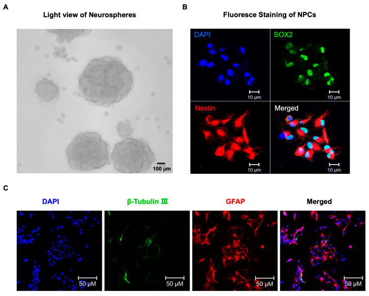
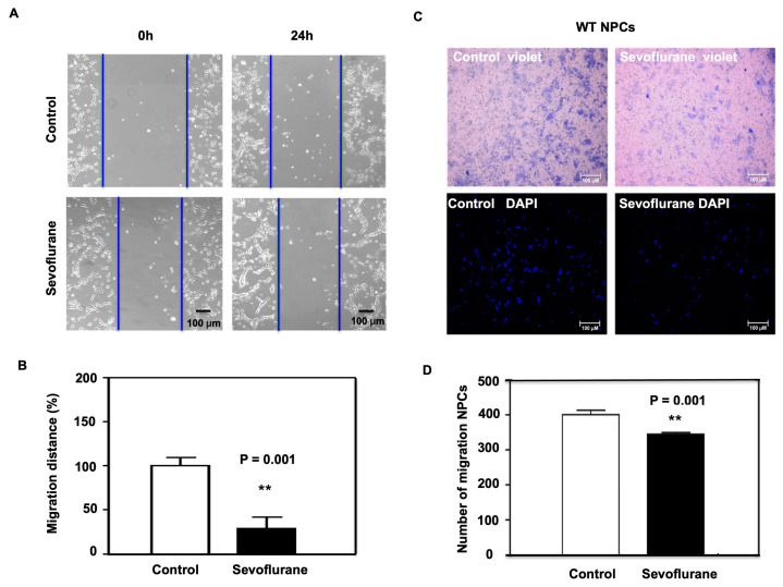
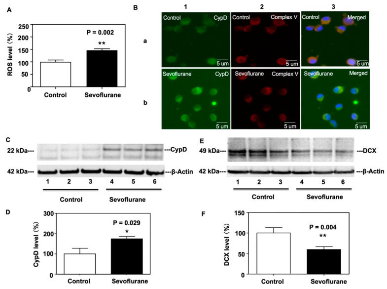
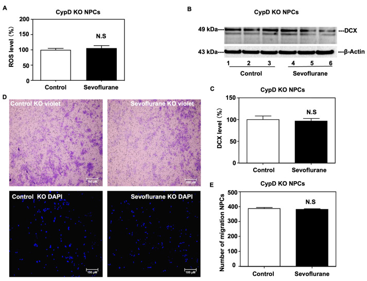
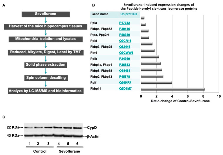
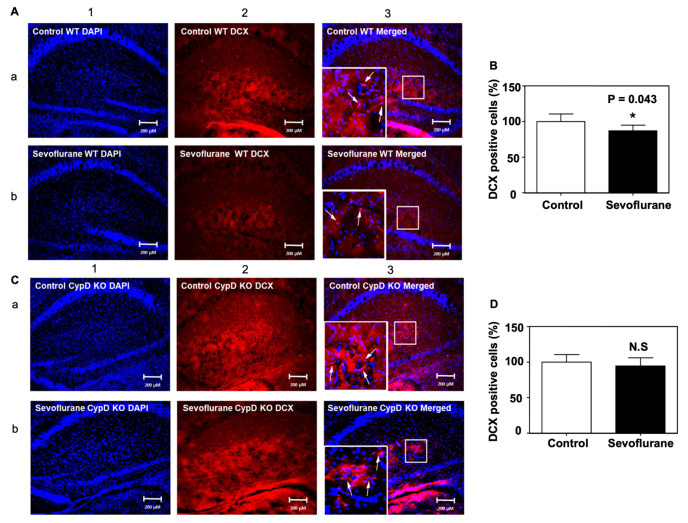
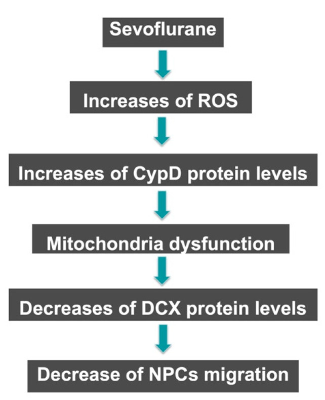
Clinical studies have suggested that repeated exposure to anesthesia and surgery at a young age may increase the risk of cognitive impairment. Our previous research has shown that sevoflurane can affect neurogenesis and cognitive function in young animals by altering cyclophilin D (CypD) levels and mitochondrial function. Neural progenitor cells (NPCs) migration is associated with cognitive function in developing brains. However, it is unclear whether sevoflurane can regulate NPCs migration via changes in CypD. To address this question, we treated NPCs harvested from wild-type (WT) and CypD knockout (KO) mice and young WT and CypD KO mice with sevoflurane. We used immunofluorescence staining, wound healing assay, transwell assay, mass spectrometry, and Western blot to assess the effects of sevoflurane on CypD, reactive oxygen species (ROS), doublecortin levels, and NPCs migration. We showed that sevoflurane increased levels of CypD and ROS, decreased levels of doublecortin, and reduced migration of NPCs harvested from WT mice in vitro and in WT young mice. KO of CypD attenuated these effects, suggesting that a sevoflurane-induced decrease in NPCs migration is dependent on CypD. Our findings have established a system for future studies aimed at exploring the impacts of sevoflurane anesthesia on the impairment of NPCs migration.

Keywords: cyclophilin D; doublecortin; migration; neural progenitor cells; sevoflurane.


Conflict of interest statement

The authors declare no conflict interest.

Figures

Figure 1
Figure 1
Identification of neural progenitor cells (NPCs). (A) Light view of NPCs, Scale bar = 100 μm. (B) Immunostaining of NPCs for DNA marker DAPI (blue), the NPCs markers SOX2 (green), and nestin (red). Scale bar = 10 μm. (C) Representative immunofluorescence images of NPCs that differentiate into neurons and astrocytes after 3 day’s differentiation culture. Double immunofluorescent staining of cells for β-tubulin III (neuronal marker, green) and GFAP (astrocyte marker, red). The nuclei are counterstained with DAPI (blue). Scale bars = 50 μm.
Figure 2
Figure 2
The effects of the sevoflurane treatment on cell migration using wound healing and transwell assays. (A) The bright-field images of the wound healing assay demonstrate cell movement 24 h after the sevoflurane treatment compared to the control condition. The blue line indicates the migration distance relative to the control condition. NPCs were maintained in the incubator between inspections. (B) Quantification of the migration shows that the sevoflurane treatment significantly reduces the migration distance in the wound healing assay compared to the control condition at 24 h after the sevoflurane treatment (** p = 0.001). (C) Transwell assay images of migrated cell stained by crystal violet (top) or DAPI (below) 24 h after the sevoflurane treatment. (D) Quantification of the transwell assay shows that the sevoflurane treatment significantly decreases the number of migrated cells as compared to the control condition (** p = 0.001; N = 10 in each group). Student’s t-test was used to analyze the data.
Figure 3
Figure 3
Sevoflurane increases ROS and CypD levels but decreases doublecortin (DCX) levels in NPCs harvested from WT mice. (A) Sevoflurane treatment (black bar) increases the levels of ROS compared to the control condition (white bar). (B) Representative images of immunofluorescent staining of CypD in NPCs harvested from WT mice. Column 1 is the CypD (green) staining, column 2 is the mitochondrial markers Complex V (red) staining, and column 3 is a merged staining image. Sevoflurane (row b) increases levels of CypD compared to the control condition (row a) in NPCs harvested from WT mice. (C) Western blot shows that the sevoflurane treatment (lanes 4 to 6) discernibly increases the levels of CypD compared to the control condition (lanes 1 to 3). There is no significant difference in β-actin levels between the sevoflurane treatment and control condition. (D) Quantitation of the Western blot illustrates increased amounts of CypD levels following the sevoflurane treatment compared to the control condition (* p = 0.029). (E) Sevoflurane treatment (lanes 4 to 6) discernibly decreases doublecortin (DCX) levels compared to the control condition. There is no significant difference in β-actin levels between the sevoflurane treatment and control condition. (F) Quantitation of the Western blot illustrates the decreased amounts of DCX levels following the sevoflurane treatment as compared to the control condition (** p = 0.004). N = 6–8 in each group. Student’s t-test was used to analyze the data.
Figure 4
Figure 4
CypD KO mitigates the sevoflurane-induced increases of ROS, decreases of DCX, and inhibition of cell migration in NPCs. (A) The sevoflurane treatment does not increase ROS levels compared to the control condition in NPCs harvested from CypD KO mice. Quantitative Western blot (B,C) analysis shows that the sevoflurane treatment does not decrease DCX levels compared to the control condition in the NPCs harvested from CypD KO mice. There is no significant difference in β-actin levels between each condition. (D) The representative transwell assay images of migrated cell stained by crystal violet (top) or DAPI (below) at 24 h after the sevoflurane treatment. (E) Quantification of the transwell assay shows that the sevoflurane treatment does not significantly affect the number of migrated NPCs harvested from CypD KO mice compared to the control condition. N = 6–8 in each group. Student’s t-test was used to analyze the data. N.S: non-significant differences.
Figure 5
Figure 5
Proteomics analyses identify mitochondrial proteins in the hippocampus of mice. (A) Experimental design and workflow for quantitative profiling of mice hippocampus mitochondrial proteomics after the sevoflurane anesthesia. (B) Quantitative proteomic analysis of the abundance ratio of Peptidyl-prolyl cis-trans isomerase (Ppif) associated proteins after the sevoflurane anesthesia. (C) Sevoflurane treatment (lanes 4 to 6) discernibly increases the levels of CypD as compared to the control condition (lanes 1 to 3) in the hippocampus of WT mice; there is no significant difference in β-actin levels between the groups.
Figure 6
Figure 6
CypD KO mitigates the sevoflurane-induced decrease of DCX-positive cells in the hippocampus of mice. (A) Representative images of immunofluorescent staining DCX in the hippocampus of WT mice. Column 1 is the DAPI (blue) nuclear staining, column 2 is the DCX (red) staining, and column 3 is the merged staining image. Sevoflurane (row b) decreases the number of DCX-positive cells compared to the control condition (row a) in the hippocampus of WT mice. (B) Quantification of the image shows that sevoflurane (black bar) significantly decreases the number of DCX-positive cells compared to the control condition (white bar) (* p = 0.043). (C) Representative images of immunofluorescent staining DCX in the hippocampus of CypD KO mice. (D) Quantification of the image shows that the sevoflurane anesthesia (black bar) does not change the number of DCX-positive cells in the hippocampus of CypD KO young mice compared to the control condition (white bar). N = 5–6 in each group. Student’s t-test was used to analyze the data. The arrows indicate the DCX positive cells in hippocampus. The Box indicates the zoom in image area in staining. N.S: non-significant differences.
Figure 7
Figure 7
Hypothetical pathway by which sevoflurane decreases mitigation. Sevoflurane triggers the production of reactive oxygen species (ROS) and increases CypD levels to induce mitochondrial dysfunction. Mitochondrial dysfunction leads to a decrease in the levels of DCX, a protein involved in neuronal migration and differentiation, resulting in a decrease in NPCs migration.

Similar articles

Cited by

References

    1. Soriano S.G., Vutskits L., Jevtovic-Todorovic V., Hemmings H.C., Davidson A., Warner D.O., Sun L.S. Thinking, fast and slow: Highlights from the 2016 BJA seminar on anaesthetic neurotoxicity and neuroplasticity. Br. J. Anaesth. 2017;119:443–447. doi: 10.1093/bja/aex238. - DOI - PMC - PubMed
    1. Wilder R.T., Flick R.P., Sprung J., Katusic S.K., Barbaresi W.J., Mickelson C., Gleich S.J., Schroeder D.R., Weaver A.L., Warner D.O. Early exposure to anesthesia and learning disabilities in a population-based birth cohort. Anesthesiology. 2009;110:796–804. doi: 10.1097/01.anes.0000344728.34332.5d. - DOI - PMC - PubMed
    1. Sun L.S., Li G., Miller T.L., Salorio C., Byrne M.W., Bellinger D.C., Ing C., Park R., Radcliffe J., Hays S.R., et al. Association Between a Single General Anesthesia Exposure Before Age 36 Months and Neurocognitive Outcomes in Later Childhood. JAMA. 2016;315:2312–2320. doi: 10.1001/jama.2016.6967. - DOI - PMC - PubMed
    1. Davidson A.J., Disma N., de Graaff J.C., Withington D.E., Dorris L., Bell G., Stargatt R., Bellinger D.C., Schuster T., Arnup S.J., et al. Neurodevelopmental outcome at 2 years of age after general anaesthesia and awake-regional anaesthesia in infancy (GAS): An international multicentre, randomised controlled trial. Lancet. 2016;387:239–250. doi: 10.1016/S0140-6736(15)00608-X. - DOI - PMC - PubMed
    1. Warner D.O., Chelonis J.J., Paule M.G., Frank R.D., Lee M., Zaccariello M.J., Katusic S.K., Schroeder D.R., Hanson A.C., Schulte P.J., et al. Performance on the Operant Test Battery in young children exposed to procedures requiring general anaesthesia: The MASK study. Br. J. Anaesth. 2019;122:470–479. doi: 10.1016/j.bja.2018.12.020. - DOI - PMC - PubMed