. 2023 Apr 4;24(7):6749.
doi: 10.3390/ijms24076749.

Dysregulation of Resolvin E1 Metabolism and Signaling in a Light-Damage Model of Age-Related Macular Degeneration

Affiliations

Dysregulation of Resolvin E1 Metabolism and Signaling in a Light-Damage Model of Age-Related Macular Degeneration

Annamaria Tisi et al. Int J Mol Sci. .

Abstract

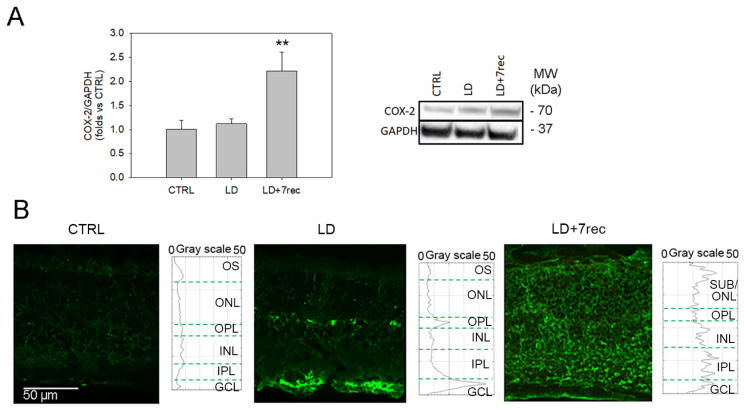
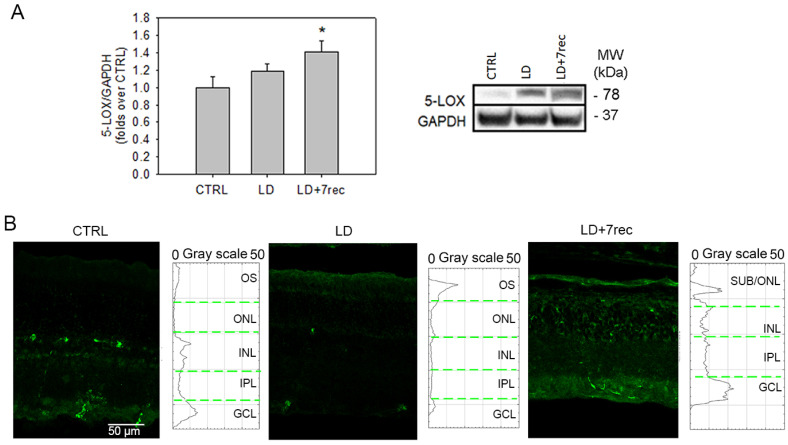
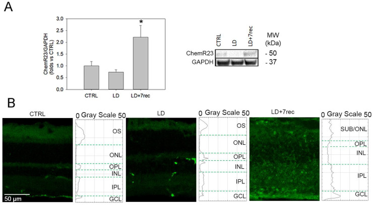
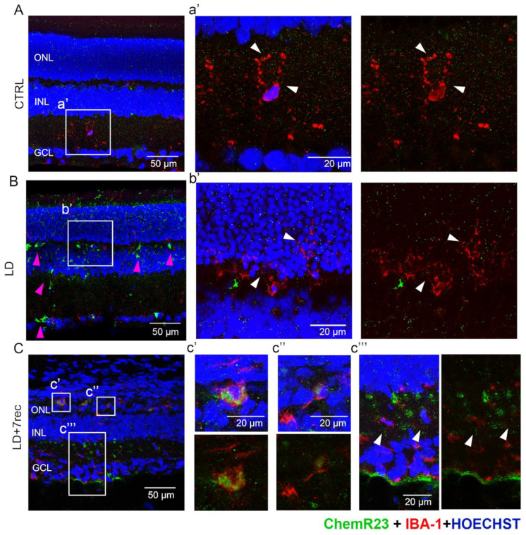
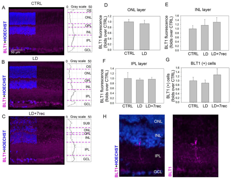
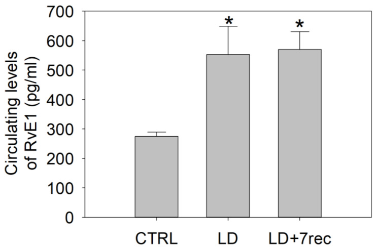
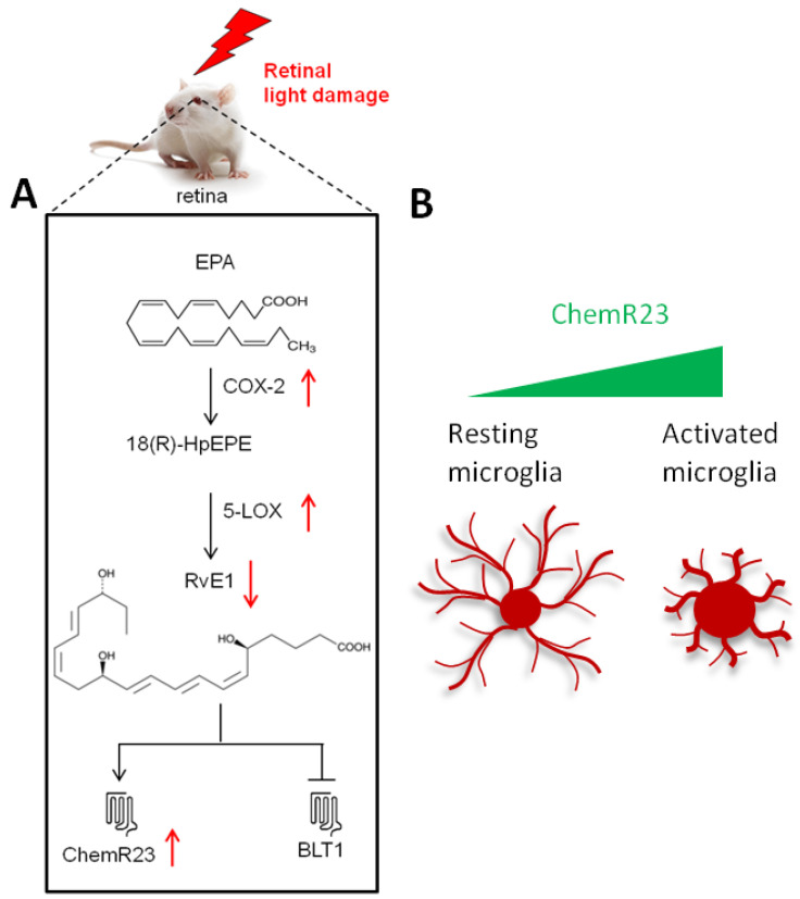
Resolvin E1 (RvE1) is an eicosapentaenoic acid-derived lipid mediator involved in the resolution of inflammation. Here, we investigated whether RvE1 alterations may occur in an animal model of age-related macular degeneration (AMD). To this end, Sprague Dawley albino rats underwent light damage (LD), and retinas and serum were analyzed immediately or seven days after treatment. Western blot of retinas showed that the RvE1 receptor ChemR23 and the RvE1 metabolic enzymes 5-LOX and COX-2 were unchanged immediately after LD, but they were significantly up-regulated seven days later. Instead, the RvE1 receptor BLT1 was not modulated by LD, and neither was the RvE1 degradative enzyme 15-PGDH. Moreover, ChemR23, 5-LOX, COX-2 and BLT1 were found to be more expressed in the inner retina under all experimental conditions, as observed through ImageJ plot profile analysis. Of note, amacrine cells highly expressed BLT1, while ChemR23 was highly expressed in the activated microglia of the outer retina. ELISA assays also showed that LD rats displayed significantly higher circulating levels and reduced retinal levels of RvE1 compared to controls. Altogether, our data indicate that RvE1 metabolism and signaling are modulated in the LD model, suggesting a potentially relevant role of this pathway in AMD.

Keywords: RvE1; SPMs; age-related macular degeneration; bioactive lipids; light damage.


Conflict of interest statement

The authors declare no conflict of interest.

Figures

Figure 1
Figure 1
COX-2 analysis. (A) Western blot analysis of COX-2 in eye cup samples of all the experimental conditions. Statistical analysis was one-way ANOVA test followed by Tukey test. Data are shown as mean ± SE (n = 6). ** p < 0.01 versus CTRL. The whole Western blot is available as Supplementary Figure S1. (B) Confocal images of retinal cryosections immunolabelled for COX-2 (green) acquired at the central dorsal area of the retina. The graphs show the plot profile fluorescence intensity of COX-2 through the retinal layers. CTRL (Control); LD (Light damage); LD + 7rec (Light damage + 7 days of recovery); OS (outer segments); ONL (Outer nuclear layer); OPL (outer plexiform layer); INL (Inner nuclear layer); IPL (inner plexiform layer); GCL (Ganglion cell layer); ONL/SUB (outer nuclear layer/subretina).
Figure 2
Figure 2
5-LOX analysis. (A) Western blot analysis of 5-LOX in eye cup samples of all the experimental conditions. Statistical analysis was a one-way ANOVA test followed by Tukey test. Data are shown as mean ± SE (n = 12). * p < 0.05 versus CTRL. The whole Western blot is available as Supplementary Figure S2. (B) Confocal images of retinal cryosections immunolabelled for 5-LOX (green) acquired at the central dorsal area of the retina. Scale bar: 50 µm. The graphs show the plot profile fluorescence intensity of 5-LOX throughout the retinal layers. CTRL (Control); LD (Light damage); LD + 7rec (Light damage + 7 days of recovery); OS (outer segments); ONL (Outer nuclear layer); OPL (outer plexiform layer); INL (Inner nuclear layer); IPL (inner plexiform layer); GCL (Ganglion cell layer).
Figure 3
Figure 3
ChemR23 analysis. (A) Western blot analysis of ChemR23 in eye cup samples of all the experimental conditions. Statistical analysis was one-way ANOVA test followed by Tukey test. Data are shown as mean ± SE (n = 6). * p < 0.05 versus CTRL. The whole Western blot is available as Supplementary Figure S4. (B) Confocal images of retinal cryosections immunolabelled for ChemR23 (green) acquired at the central dorsal area of the retina. Scale bar: 50 µm. The graphs show the plot profile fluorescence intensity of ChemR23 throughout the retinal layers. CTRL (Control); LD (Light damage); LD + 7rec (Light damage + 7 days of recovery); OS (outer segments); ONL (Outer nuclear layer); OPL (outer plexiform layer); INL (Inner nuclear layer); IPL (inner plexiform layer); GCL (Ganglion cell layer).
Figure 4
Figure 4
Double immunostaining for IBA-1 and ChemR23. (AC) Confocal images of retinal cryosections labelled for IBA-1 (red) and ChemR23 (green), and counterstained with bisbenzimide nuclear dye (blue) of (A) CTRL, (B) LD and (C) LD + 7rec retinas. Scale bar: 50 µm. High magnifications (a’,b’,c’,c’’,c’’’), scale bar 20 µm. The white arrows show detailed localization of ChemR23 in IBA-1 (+) cells. The pink arrows indicate increased ChemR23 signal in the LD group not co-localizing with IBA-1 (+) cells. CTRL (Control); LD (Light damage); LD + 7rec (Light damage + 7 days of recovery); ONL (Outer nuclear layer); INL (Inner nuclear layer); GCL (Ganglion cell layer).
Figure 5
Figure 5
BLT1 analysis. (AC) Confocal images and plot profile of fluorescence intensity of retinal cryosections from CTRL, LD and LD + 7rec groups, respectively, immunolabelled for BLT1 (pink). Nuclei were stained with Bisbenzimide (blue). The images refer to the central dorsal area of the retina. Scale bar: 50 µm. BLT1 fluorescence intensity in (D) the ONL; (E) the INL; (F) in the IPL. (G) Quantitative analysis of BLT1 (+) cells from the superior to the inferior edge of retinal cryosections. Data are shown as mean ± SE (n = 4). (H) High magnification of random BLT1 (+) cells (pink). CTRL (Control); LD (Light damage); LD + 7rec (Light damage + 7 days of recovery); OS (outer segments); ONL (Outer nuclear layer); OPL (outer plexiform layer); INL (Inner nuclear layer); IPL (inner plexiform layer); GCL (Ganglion cell layer).
Figure 6
Figure 6
Quantification of RvE1 in the serum of LD rats. ELISA assay was performed on serum samples from all experimental groups. Statistical analysis was performed by one-way ANOVA test followed by Tukey test. Data are shown as mean ± SE (n = 4). * p < 0.05 versus CTRL. CTRL (Control); LD (Light damage); LD + 7rec (Light damage + 7 days of recovery).
Figure 7
Figure 7
Schematic illustration of the main findings of the study. The scheme summarizes the results obtained from the retinas 7 days after LD (LD + 7rec group). (A) Effects of light damage (LD) on RvE1 metabolism and signaling in the rat retinas; the black lines indicate RvE1 metabolism and signaling, while the red arrows indicate the events induced by LD. (B) Schematic cartoon illustrating the different expressions observed in retinal resting and activated microglia.

Similar articles

Cited by

References

    1. Serhan C.N. Pro-resolving lipid mediators are leads for resolution physiology. Nature. 2014;510:92–101. doi: 10.1038/nature13479. - DOI - PMC - PubMed
    1. Leuti A., Fazio D., Fava M., Piccoli A., Oddi S., Maccarrone M. Bioactive lipids, inflammation and chronic diseases. Adv. Drug Deliv. Rev. 2020;159:133–169. doi: 10.1016/j.addr.2020.06.028. - DOI - PubMed
    1. Serhan C.N., Hong S., Gronert K., Colgan S.P., Devchand P.R., Mirick G., Moussignac R.L. Resolvins: A family of bioactive products of omega-3 fatty acid transformation circuits initiated by aspirin treatment that counter proinflammation signals. J. Exp. Med. 2002;196:1025–1037. doi: 10.1084/jem.20020760. - DOI - PMC - PubMed
    1. Trotta M.C., Gesualdo C., Petrillo F., Lepre C.C., Della Corte A., Cavasso G., Maggiore G., Hermenean A., Simonelli F., D’amico M., et al. Resolution of Inflammation in Retinal Disorders: Briefly the State. Int. J. Mol. Sci. 2022;23:4501. doi: 10.3390/ijms23094501. - DOI - PMC - PubMed
    1. Rossi S., Di Filippo C., Gesualdo C., Potenza N., Russo A., Trotta M.C., Zippo M.V., Maisto R., Ferraraccio F., Simonelli F., et al. Protection from endotoxic uveitis by intravitreal resolvin D1: Involvement of lymphocytes, miRNAs, ubiquitin-proteasome, and M1/M2 macrophages. Mediat. Inflamm. 2015;2015:149381. doi: 10.1155/2015/149381. - DOI - PMC - PubMed

Substances