

The Different Pathways of Epicardial Adipose Tissue across the Heart Failure Phenotypes: From Pathophysiology to Therapeutic Target
- PMID: 37047810
- PMCID: PMC10095298
- DOI: 10.3390/ijms24076838
The Different Pathways of Epicardial Adipose Tissue across the Heart Failure Phenotypes: From Pathophysiology to Therapeutic Target
Abstract
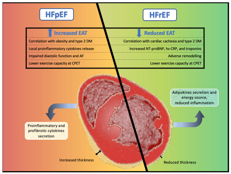
Epicardial adipose tissue (EAT) is an endocrine and paracrine organ constituted by a layer of adipose tissue directly located between the myocardium and visceral pericardium. Under physiological conditions, EAT exerts protective effects of brown-like fat characteristics, metabolizing excess fatty acids, and secreting anti-inflammatory and anti-fibrotic cytokines. In certain pathological conditions, EAT acquires a proatherogenic transcriptional profile resulting in increased synthesis of biologically active adipocytokines with proinflammatory properties, promoting oxidative stress, and finally causing endothelial damage. The role of EAT in heart failure (HF) has been mainly limited to HF with preserved ejection fraction (HFpEF) and related to the HFpEF obese phenotype. In HFpEF, EAT seems to acquire a proinflammatory profile and higher EAT values have been related to worse outcomes. Less data are available about the role of EAT in HF with reduced ejection fraction (HFrEF). Conversely, in HFrEF, EAT seems to play a nutritive role and lower values may correspond to the expression of a catabolic, adverse phenotype. As of now, there is evidence that the beneficial systemic cardiovascular effects of sodium-glucose cotransporter-2 receptors-inhibitors (SGLT2-i) might be partially mediated by inducing favorable modifications on EAT. As such, EAT may represent a promising target organ for the development of new drugs to improve cardiovascular prognosis. Thus, an approach based on detailed phenotyping of cardiac structural alterations and distinctive biomolecular pathways may change the current scenario, leading towards a precision medicine model with specific therapeutic targets considering different individual profiles. The aim of this review is to summarize the current knowledge about the biomolecular pathway of EAT in HF across the whole spectrum of ejection fraction, and to describe the potential of EAT as a therapeutic target in HF.
Keywords: biomolecular pathways; cardiovascular disease; epicardial adipose tissue; heart failure.
Conflict of interest statement
The authors declare no conflict of interest.
Figures
Similar articles
-
Epicardial Adipose Tissue and Heart Failure, Friend or Foe?Diabetes Metab J. 2024 May;48(3):373-384. doi: 10.4093/dmj.2023.0190. Epub 2024 Feb 2. Diabetes Metab J. 2024. PMID: 38310880 Free PMC article. Review.
-
Drugs That Ameliorate Epicardial Adipose Tissue Inflammation May Have Discordant Effects in Heart Failure With a Preserved Ejection Fraction as Compared With a Reduced Ejection Fraction.J Card Fail. 2019 Dec;25(12):986-1003. doi: 10.1016/j.cardfail.2019.09.002. Epub 2019 Sep 18. J Card Fail. 2019. PMID: 31541742 Review.
-
Role of epicardial adipose tissue in heart failure with preserved ejection fraction: An emerging molecular mechanism and therapeutic potential.Obes Rev. 2025 Jul;26(7):e13912. doi: 10.1111/obr.13912. Epub 2025 Mar 1. Obes Rev. 2025. PMID: 40022641 Free PMC article. Review.
-
Diverging role of epicardial adipose tissue across the entire heart failure spectrum.ESC Heart Fail. 2023 Dec;10(6):3419-3429. doi: 10.1002/ehf2.14483. Epub 2023 Sep 11. ESC Heart Fail. 2023. PMID: 37697706 Free PMC article.
-
Pericardium, epicardial adipose tissue, and heart failure with preserved ejection fraction: Pathophysiology, quantification and treatment target.Int J Cardiol. 2024 Oct 1;412:132303. doi: 10.1016/j.ijcard.2024.132303. Epub 2024 Jun 27. Int J Cardiol. 2024. PMID: 38944349 Review.
Cited by
-
Targeting Epicardial/Pericardial Adipose Tissue in Cardiovascular Diseases: A Novel Therapeutic Strategy.Rev Cardiovasc Med. 2025 Mar 13;26(3):26128. doi: 10.31083/RCM26128. eCollection 2025 Mar. Rev Cardiovasc Med. 2025. PMID: 40160564 Free PMC article. Review.
-
Sodium-glucose cotransporter 2 inhibitors induce anti-inflammatory and anti-ferroptotic shift in epicardial adipose tissue of subjects with severe heart failure.Cardiovasc Diabetol. 2024 Jun 28;23(1):223. doi: 10.1186/s12933-024-02298-9. Cardiovasc Diabetol. 2024. PMID: 38943140 Free PMC article.
-
Epicardial adipose tissue volume and density are associated with heart failure with improved ejection fraction.Cardiovasc Diabetol. 2024 Aug 3;23(1):283. doi: 10.1186/s12933-024-02376-y. Cardiovasc Diabetol. 2024. PMID: 39097703 Free PMC article.
-
Open-bore MRI Scanner Assessment of Epicardial Adipose Tissue after Bariatric Surgery: A Pilot Study.Endocr Metab Immune Disord Drug Targets. 2025;25(2):173-188. doi: 10.2174/0118715303310680240607114244. Endocr Metab Immune Disord Drug Targets. 2025. PMID: 39171595 Free PMC article.
-
Association of Epicardial Adipose Tissue with Novel Inflammation and Heart Failure Biomarkers in Type 2 Diabetes Patients: Effect of Metabolic Control.J Clin Med. 2025 Jul 2;14(13):4687. doi: 10.3390/jcm14134687. J Clin Med. 2025. PMID: 40649060 Free PMC article.
References
-
- Janovska P., Melenovsky V., Svobodova M., Havlenova T., Kratochvilova H., Haluzik M., Hoskova E., Pelikanova T., Kautzner J., Monzo L., et al. Dysregulation of epicardial adipose tissue in cachexia due to heart failure: The role of natriuretic peptides and cardiolipin. J. Cachexia Sarcopenia Muscle. 2020;11:1614–1627. doi: 10.1002/jcsm.12631. - DOI - PMC - PubMed
-
- Parisi V., Rengo G., Pagano G., D’Esposito V., Passaretti F., Caruso A., Grimaldi M.G., Lonobile T., Baldascino F., De Bellis A., et al. Epicardial adipose tissue has an increased thickness and is a source of inflammatory mediators in patients with calcific aortic stenosis. Int. J. Cardiol. 2015;186:167–169. doi: 10.1016/j.ijcard.2015.03.201. - DOI - PubMed
Publication types
MeSH terms
LinkOut - more resources
Full Text Sources
Medical
Research Materials
Miscellaneous
