

Salvianolic-Acid-B-Loaded HA Self-Healing Hydrogel Promotes Diabetic Wound Healing through Promotion of Anti-Inflammation and Angiogenesis
- PMID: 37047818
- PMCID: PMC10095058
- DOI: 10.3390/ijms24076844
Salvianolic-Acid-B-Loaded HA Self-Healing Hydrogel Promotes Diabetic Wound Healing through Promotion of Anti-Inflammation and Angiogenesis
Abstract
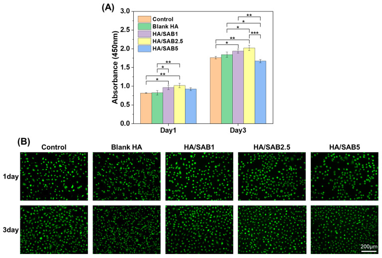
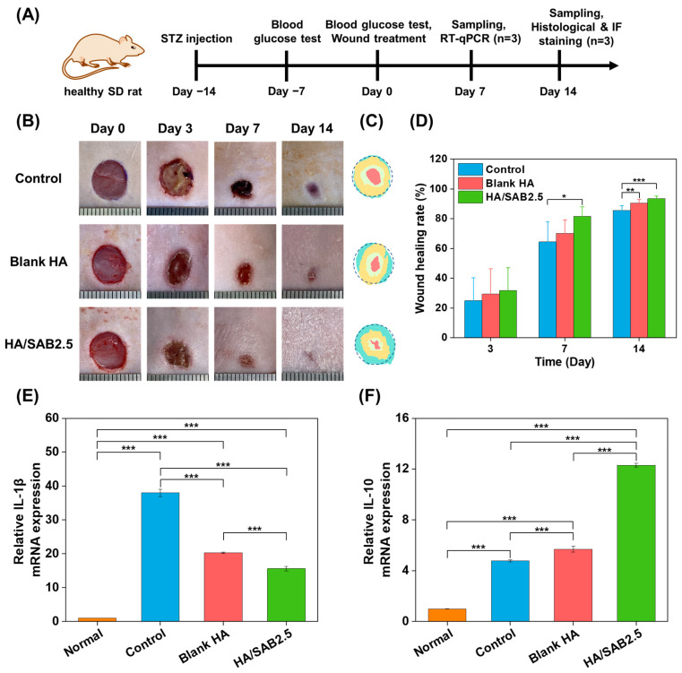
Inflammatory dysfunction and angiogenesis inhibition are two main factors leading to the delayed healing of diabetic wounds. Hydrogels with anti-inflammatory and angiogenesis-promoting effects have been considered as promising wound care materials. Herein, a salvianolic acid B (SAB)-loaded hyaluronic acid (HA) self-healing hydrogel (HA/SAB) with anti-inflammatory and pro-angiogenesis capacities for diabetic wound healing is reported. The HA hydrogel was prepared via the covalent cross-linking of aldehyde groups in oxidized HA (OHA) and hydrazide groups in adipic dihydrazide (ADH)-modified HA (HA-ADH) with the formation of reversible acylhydrazone bonds. The obtained HA hydrogel exhibited multiple favorable properties such as porous structures, excellent self-healing properties, a sustainable release capacity of SAB, as well as excellent cytocompatibility. In addition, the effects of the SAB-loaded HA self-healing hydrogel were investigated via a full-thickness skin defect model using diabetic rats. The HA/SAB hydrogel showed enhanced skin regeneration effects with accelerated wound closure, shorter remaining dermal space length, thicker granulation tissue formation, and more collagen deposition. Furthermore, reduced inflammatory response and enhanced vascularization were found with HA/SAB2.5 hydrogel-treated wounds, indicating that the hydrogel promotes diabetic wound healing through the promotion of anti-inflammation and angiogenesis. Our results suggest that the fabricated SAB-loaded HA self-healing hydrogel is promising as a wound dressing for the treatment of diabetic wounds.
Keywords: angiogenesis; anti-inflammation; diabetic wound healing; salvianolic acid B; self-healing hydrogel.
Conflict of interest statement
The authors declare no conflict of interest.
Figures


Similar articles
-
Promote anti-inflammatory and angiogenesis using a hyaluronic acid-based hydrogel with miRNA-laden nanoparticles for chronic diabetic wound treatment.Int J Biol Macromol. 2021 Jan 1;166:166-178. doi: 10.1016/j.ijbiomac.2020.10.129. Epub 2020 Oct 22. Int J Biol Macromol. 2021. PMID: 33172616
-
A hyaluronic acid hydrogel as a mild photothermal antibacterial, antioxidant, and nitric oxide release platform for diabetic wound healing.J Control Release. 2024 Jun;370:543-555. doi: 10.1016/j.jconrel.2024.05.011. Epub 2024 May 10. J Control Release. 2024. PMID: 38729434
-
An Injectable Ibuprofen Sustained-Release Composite Hydrogel System Effectively Accelerates Diabetic Wound Healing via Anti-Inflammatory Effects and Angiogenesis.Int J Nanomedicine. 2025 Apr 11;20:4535-4550. doi: 10.2147/IJN.S504924. eCollection 2025. Int J Nanomedicine. 2025. PMID: 40236520 Free PMC article.
-
Development of hyaluronic acid-based hydrogels for chronic diabetic wound healing: A review.Int J Biol Macromol. 2025 May;308(Pt 1):142273. doi: 10.1016/j.ijbiomac.2025.142273. Epub 2025 Mar 18. Int J Biol Macromol. 2025. PMID: 40112998 Review.
-
Hyaluronic Acid-Based Self-Healing Hydrogels for Diabetic Wound Healing.Adv Healthc Mater. 2025 Feb;14(4):e2404255. doi: 10.1002/adhm.202404255. Epub 2024 Dec 25. Adv Healthc Mater. 2025. PMID: 39722163 Review.
Cited by
-
Unveiling the therapeutic potential of medicinal plants in zebrafish caudal fin regeneration and wound healing: a systematic review.Fish Physiol Biochem. 2025 Apr 11;51(2):80. doi: 10.1007/s10695-025-01495-x. Fish Physiol Biochem. 2025. PMID: 40214856
-
Injectable Hydrogels Based on Hyaluronic Acid and Gelatin Combined with Salvianolic Acid B and Vascular Endothelial Growth Factor for Treatment of Traumatic Brain Injury in Mice.Molecules. 2024 Apr 10;29(8):1705. doi: 10.3390/molecules29081705. Molecules. 2024. PMID: 38675525 Free PMC article.
-
Advancements and Challenges in Self-Healing Hydrogels for Wound Care.Gels. 2024 Apr 1;10(4):241. doi: 10.3390/gels10040241. Gels. 2024. PMID: 38667660 Free PMC article. Review.
References
-
- Nguyen T.T., Jones J.I., Wolter W.R., Perez R.L., Schroeder V.A., Champion M.M., Hesek D., Lee M., Suckow M.A., Mobashery S., et al. Hyperbaric oxygen therapy accelerates wound healing in diabetic mice by decreasing active matrix metalloproteinase-9. Wound Repair Regen. 2020;28:194–201. doi: 10.1111/wrr.12782. - DOI - PubMed
MeSH terms
Substances
Grants and funding
- LQ21H180004/Zhejiang Provincial Natural Science Foundation of China
- 81930111/National Natural Science Foundation of China
- 2022TS002/College level scientific research cultivation project of Zhejiang Chinese Medical University
- 2022JKZKTS20 2021RCZXZK23 2022GJYY025/Research Project of Zhejiang Chinese Medical University
- 2023ZF013 2023ZF157/Research Project on Chinese Medicine Health Services
LinkOut - more resources
Full Text Sources
