Review
. 2023 Mar 23;12(7):986.
doi: 10.3390/cells12070986.

Substances of Natural Origin in Medicine: Plants vs. Cancer

Affiliations
Review

Substances of Natural Origin in Medicine: Plants vs. Cancer

Adrianna Gielecińska et al. Cells. .

Abstract

Continuous monitoring of the population's health is the main method of learning about disease prevalence. National and international data draw attention to the persistently high rates of cancer incidence. This necessitates the intensification of efforts aimed at developing new, more effective chemotherapeutic and chemopreventive drugs. Plants represent an invaluable source of natural substances with versatile medicinal properties. Multidirectional activities exhibited by natural substances and their ability to modulate key signaling pathways, mainly related to cancer cell death, make these substances an important research direction. This review summarizes the information regarding plant-derived chemotherapeutic drugs, including their mechanisms of action, with a special focus on selected anti-cancer drugs (paclitaxel, irinotecan) approved in clinical practice. It also presents promising plant-based drug candidates currently being tested in clinical and preclinical trials (betulinic acid, resveratrol, and roburic acid).

Keywords: betulinic acid; cancer; irinotecan; paclitaxel; resveratrol; roburic acid.


Conflict of interest statement

The authors declare no conflict of interest.

Figures
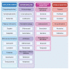
Figure 1
Figure 1
The main types of chemotherapeutics sorted by their mechanism of action. Among the most popular are alkylating compounds, antimetabolites, topoisomerase inhibitors, and mitotic spindle inhibitors. Created with BioRender.com.
Figure 2
Figure 2
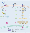
The activation of nuclear factor kappa-light-chain-enhancer of activated B cells (NF-κB) and the extracellular signal-regulated kinase (ERK)/mitogen-activated protein kinase (MAPK) and phosphoinositide 3-kinase (PI3K)/protein kinase B (AKT) pathways that support the development of the tumor and the pro-inflammatory cellular environment. Phosphorylation of B-cells inhibitor (IκB) by IκB kinase (IKKβ) as a result of cellular signals (growth factors, cytokines) received by receptor (tumor necrosis factor receptors (TNFR)), results in heterodimer disconnection, release of NF-κB, and degradation of IκB complex component. The released NF-κB translocates from the cytoplasm to the nucleus, where it acts as a transcription factor responsible for the expression of proteins involved in cell proliferation, migration, and inflammation. The activation of receptor tyrosine kinases (RTKs) initiates activation of ERK/MAPK and PI3K/AKT pathways, promoting survival, migration, and invasion of cancer cells. Binding of the ligand to the RTKs causes the “turning on” of the rat sarcoma virus (RAS) protein through GDP-to-GTP exchange, which activates the RAF proto-oncogene serine/threonine-protein kinase (RAF), mitogen-activated protein kinase (MEK) and ERK kinase, leading to the growth of cancer cells and supporting the transcription of pro-inflammatory factors. Similarly, PI3K is activated via RTKs, which catalyze the phosphorylation of phosphatidylinositol 4,5-biphosphate (PIP2) to phosphatidylinositol (3,4,5)-triphosphate (PIP3). As a result of the interaction of PIP3 and phosphatidylinositol kinase (PDK-1), AKT is activated. Being a serine-threonine kinase, AKT phosphorylates numerous proteins involved in the regulation of cell proliferation, growth, and survival. The key substrates for AKT are the mammalian target of rapamycin (mTOR) and the pro-apoptotic factor BCL-2-associated death promoter (BAD), which are capable of inhibiting apoptosis, and nuclear transcription factors fork head box O (FOXO). The use of betulinic acid (BA) prevents the phosphorylation of IκB, and thus, the release and translocation of NF-κB into the nucleus. As a consequence, the expression of inflammatory mediators and enzymes is not induced. BA also inhibits the activation of ERK/MAPK and PI3K/AKT signaling pathways, suppressing mechanisms oriented toward tumor growth and development. Created with BioRender.com.
Figure 3
Figure 3
Tumor necrosis factor α (TNF-α) and vascular endothelial growth factor (VEGF) with pro-angiogenic function are overexpressed in tumors depending on transcription factors, i.e., transcription factor Sp1 (SP1) and transcription factor Sp3 (SP3). They are supported by the nuclear factor kappa-light-chain-enhancer of activated B cells (NF-κB), which bind to specific sequences known as NF-κB response elements (NRE) and GC-BOXes. The presence of VEGF in the cell enhances the activation of the rat sarcoma virus-GTP (RAS-GTP)/RAF proto-oncogene serine/threonine-protein kinase (RAF)/mitogen-activated protein kinase (MEK)/extracellular signal-regulated kinase (ERK) and phosphoinositide 3-kinase (PI3K)/phosphatidylinositol kinase (PDK-1)/protein kinase B (AKT) pathways, promoting invasive potential and tumor metastases. Betulinic acid (BA) prevents the translocation of NF-κB to the nucleus and promotes the degradation of SP1 and SP3, down-regulating VEGF. Created with BioRender.com.
Figure 4
Figure 4
The process of autophagolysosome formation. Stimulated by the mitogen-activated protein kinase (MAPK) and protein kinase B (AKT) pathways, mammalian target of rapamycin (mTOR) and 5’ AMP-activated protein kinase (AMPK) regulate autophagy by interacting with Unc-51 like autophagy activating kinase (ULK1) complex and autophagy-related protein 13. ULK1, activated by AMPK, initiates autophagosome formation from intracellular membranes and its elongation. Then the autophagosome enters the elongation phase. Under the influence of the ATG3, ATG4 and ATG7 proteins, protein light chain (LC3) protein is converted to the LC3-I, LC3-II and LC3-III forms. The autophagy-related proteins (ATGs) (like ATG5, ATG7, ATG10, ATG12, ATG16) together with LC3-III contribute to the maturation of autophagosome that, through fusion with the lysosome, forms an autophagolysosome, which is a structure capable of enzymatic digestion of proteins. Created with BioRender.com.
Figure 5
Figure 5
(A) Resveratrol can induce DNA damage, resulting in the recruitment of ataxia teleangiectasia mutated (ATM)/ataxia teleangiectasia and RAD3 related (ATR) kinases that phosphorylate checkpoint kinase 1 (CHK1)/checkpoint kinase 2 (CHK2) serine-threonine kinases. This prevents the interaction of tumor protein P53 (TP53) with mouse homolog of double minute 2 (MDM2), thereby protecting the TP53 protein from degradation. TP53 induces the expression of genes involved in cell cycle arrest and apoptosis. (B) Epithelial–mesenchymal transition (EMT) is initiated by many factors, including the activation of transforming growth factor β receptor (TGF-βR) by the native ligand transforming growth factor β (TGF-β). This event initiates the signaling through rat sarcoma virus-GTP (RAS-GTP)/RAF proto-oncogene serine/threonine-protein kinase (RAF)/mitogen-activated protein kinase (MEK)/extracellular signal-regulated kinase (ERK) and phosphoinositide 3-kinase (PI3K)/phosphatidylinositol kinase (PDK-1)/protein kinase B (AKT)/mammalian target of rapamycin (mTOR)/ nuclear factor kappa-light-chain-enhancer of activated B Cells (NF-κB) pathways, resulting in the expression of EMT-related factors. EMT can also be promoted by c-Jun N-terminal kinase (JNK) and p38 kinase, which are members of the mitogen-activated protein kinase (MAPK) pathway. They are activated by MAP kinase (MKK) and TNF receptor-associated factor 6 (TRAF6) with K63-linked polyubiquitin chains by TGF-β-dependent polyubiquitinylation. Resveratrol inhibits TGF-β and TRAF6, preventing EMT of cancer cells. Created with BioRender.com.

Similar articles

Cited by

References

    1. Fitzmaurice C., Allen C., Barber R.M., Barregard L., Bhutta Z.A., Brenner H., Dicker D.J., Chimed-Orchir O., Dandona R., Dandona L., et al. Global, Regional, and National Cancer Incidence, Mortality, Years of Life Lost, Years Lived with Disability, and Disability-Adjusted Life-Years for 32 Cancer Groups, 1990 to 2015: A Systematic Analysis for the Global Burden of Disease Study. JAMA Oncol. 2017;3:524–548. doi: 10.1001/jamaoncol.2016.5688. - DOI - PMC - PubMed
    1. Kocarnik J.M., Compton K., Dean F.E., Fu W., Gaw B.L., Harvey J.D., Henrikson H.J., Lu D., Pennini A., Xu R., et al. Cancer Incidence, Mortality, Years of Life Lost, Years Lived With Disability, and Disability-Adjusted Life Years for 29 Cancer Groups from 2010 to 2019. JAMA Oncol. 2022;8:420–444. doi: 10.1001/jamaoncol.2021.6987. - DOI - PMC - PubMed
    1. Newman D.J., Cragg G.M. Natural Products as Sources of New Drugs over the Nearly Four Decades from 01/1981 to 09/2019. J. Nat. Prod. 2020;83:770–803. doi: 10.1021/acs.jnatprod.9b01285. - DOI - PubMed
    1. Xu Z., Eichler B., Klausner E.A., Duffy-Matzner J., Zheng W. Lead/Drug Discovery from Natural Resources. Molecules. 2022;27:8280. doi: 10.3390/molecules27238280. - DOI - PMC - PubMed
    1. Deng L.-J., Qi M., Li N., Lei Y.-H., Zhang D.-M., Chen J.-X. Natural Products and Their Derivatives: Promising Modulators of Tumor Immunotherapy. J. Leukoc. Biol. 2020;108:493–508. doi: 10.1002/JLB.3MR0320-444R. - DOI - PMC - PubMed