. 2023 Mar 23;12(7):989.
doi: 10.3390/cells12070989.

Retinal Development in a Precocial Bird Species, the Quail (Coturnix coturnix, Linnaeus 1758)

Affiliations

Retinal Development in a Precocial Bird Species, the Quail (Coturnix coturnix, Linnaeus 1758)

Guadalupe Álvarez-Hernán et al. Cells. .

Abstract

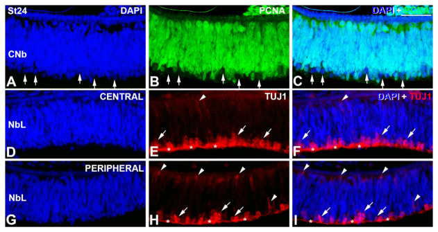
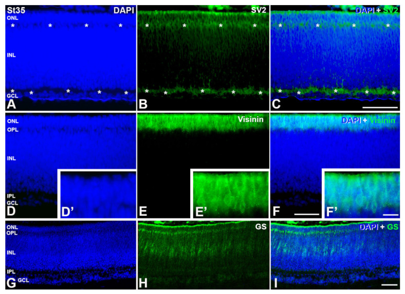
The quail (Coturnix coturnix, Linnaeus 1758), a notable model used in developmental biology, is a precocial bird species in which the processes of retinal cell differentiation and retinal histogenesis have been poorly studied. The purpose of the present research is to examine the retinogenesis in this bird species immunohistochemically and compare the results with those from previous studies in precocial and altricial birds. We found that the first PCNA-negative nuclei are detected at Stage (St) 21 in the vitreal region of the neuroblastic layer, coinciding topographically with the first αTubAc-/Tuj1-/Isl1-immunoreactive differentiating ganglion cells. At St28, the first Prox1-immunoreactive nuclei can be distinguished in the vitreal side of the neuroblastic layer (NbL), but also the first visinin-immunoreactive photoreceptors in the scleral surface. The inner plexiform layer (IPL) emerges at St32, and the outer plexiform layer (OPL) becomes visible at St35-the stage in which the first GS-immunoreactive Müller cells are distinguishable. Newly hatched animals show a well-developed stratified retina in which the PCNA-and pHisH3-immunoreactivies are absent. Therefore, retinal cell differentiation in the quail progresses in the stereotyped order conserved among vertebrates, in which ganglion cells initially appear and are followed by amacrine cells, horizontal cells, and photoreceptors. Müller glia are one of the last cell types to be born. Plexiform layers emerge following a vitreal-to-scleral gradient. Finally, our results suggest that there are no significant differences in the timing of different events involved in retinal maturation between the quail and the chicken, but the same events are delayed in an altricial bird species.

Keywords: altricial; cell differentiation; immunohistochemistry; precocial; quail; retina; retinogenesis.


Conflict of interest statement

The authors declare no conflict of interest.

Figures

Figure 1
Figure 1
Onset of cell differentiation in the retina of C. corturnix at St 21- of development. DAPI staining reveals that quail retina is composed of a NbL at this stage (A,E,I,L). The first α-Tub-positive cells are detected at this stage in the inner surface of the neural tissue (arrows in B,D,F,H). Radially oriented α-Tub-immunoreactive elements are also observed (B,D,F,H). pHisH3 antibody shows abundant mitotic cells distributed throughout the scleral surface of the NbL (arrowheads in (C,D,G,H)). Isl1-immunoreactivity is observed in cell nuclei located in the inner surface of the NbL. These Isl1-positive nuclei are restricted to the central region of the undifferentiated retina (arrows in (J,K,M,N)). L, lens; NbL, neuroblastic layer. Scale bars: 100 μm.
Figure 2
Figure 2
PCNA (AC) and TUJ1 (DI) immunoreactivity in retinal cryosections of C. coturnix at St 24–25 of development. DAPI staining reveals that quail retina is composed of a NbL at this stage (A,D,G). PCNA-negative nuclei point to those cells that have started the differentiation process (arrows in (AC)). Central (DF) and peripheral (GI) regions of the undifferentiated retina show strong TUJ1-immunoreactivity in both migratory neuroblasts (arrowheads in (E,F,H,I)), and ganglion cell axons in the optic fiber layer (asterisks). Notice that the number of TUJ1 immunoreactive elements located in the inner surface seems to be higher in the central region. NbL, neuroblastic layer. Scale bars: 100 μm.
Figure 3
Figure 3
Detection of markers of cell differentiation in retinal cryosections of C. coturnix at St28 of development. DAPI staining reveals that quail retina is composed of a NbL at this stage (A,D,H,K,O,R). PCNA-immunonegative nuclei are found in the inner surface of the NbL (arrowheads in (AC, A′C′), but also in the outer surface of the undifferentiated retina (double arrowheads in (AC)). Strong α-Tub-immunoreactivity is detected in the presumptive OFL (asterisks in (E,G)) and in the cytoplasm of migratory neuroblasts (double arrowheads in (E,G)). pHisH3 immunoreactive mitotic figures are abundant in the scleral surface of the NbL (arrowheads in (F,G)). Islet1 immunosignal is located in both the nuclei of migratory neuroblasts (arrowheads in (I,J)) and cell nuclei located in the inner surface of the NbL (arrows in (I,J)). Double immunostaining reveals that some TUJ1 immunoreactive cells located in the inner surface of the NbL also express CR (arrowheads in (LN) and (K’N’)). The presumptive OFL is also strongly TUJ1-immunoreactive (asterisks in M,N). Prox1 is first detected at this stage in cell nuclei located close to the vitreal surface of the NbL (arrows in (P,Q)). Sparsely visinin-immunoreactive cells are firstly distinguishable in the outer surface of the NbL (arrows in (S,T)). NbL, neuroblastic layer. Scale bars: 100 μm in A-T; 30 μm in (A′,C′,K′N′).
Figure 4
Figure 4
Immunodetection of different cell markers in retinal cryosections of C. coturnix at St32 of development. DAPI staining reveals an incipient emergence of the IPL (A,E,H,K,N). A faint immunostaining against SV2 is detected in the developing IPL (asterisks in (B,C)). Most of the cells located in the GCL are doubly labeled for CR and TUJ1 (arrows in (DG)). Single TUJ1-labeled cells are also detected in the pGCL (arrowheads in (F,G)) and in more external regions (double arrowheads in (F,G)). A few rows of Isl1-positive nuclei can be observed in the pGCL (I,J). Sparse Isl1-immunopositive nuclei are also observed in more external regions (arrowheads in (I,J)). Prox1-immunostaining localizes in the innermost region of the oNbL and in the GCL (arrows in (L,M)), but also in more external regions of the oNbL (double arrowheads). Visinin-immunoreactive cells are mainly located in the more external region of the oNbL, but sparse labeled cells can be distinguished in more internal regions (arrowheads in (O,P)). Visinin-positive cells clearly show a cell somata with a short external process (O′,P′). GCL, ganglion cell layer; IPL, inner plexiform layer; NbL, neuroblastic layer; oNbL, outer neuroblastic layer; pGCL, presumptive ganglion cell layer. Scale bars: 100 μm in (AP); 30 μm in (N′P′).
Figure 5
Figure 5
Cell markers in retinal cryosections of C. coturnix at St35 of development. DAPI staining reveals the multilayered structure of the quail retina (A,E,I). In the central retina, PCNA-positive proliferating cells are mainly restricted to the outer half of the INL (B,D). pHisH3-immunoreactive mitotic figures are sparse and are mainly found in the outer INL and in the ONL (arrowheads in (C,D)). Abundant ganglion and amacrine cells are immunoreactive against CR (F,H) and TUJ1 (G,H). A few ganglion cells co-express these markers (F′H′). Islet1-immunoreactivity identify ganglion, amacrine and horizontal cells (J,L). Prox1-positive nuclei are found in the presumptive horizontal cell layer (K,L) and some of them also express Isl1 (J′L′). GCL, ganglion cell layer; INL, inner nuclear layer; IPL, inner plexiform layer; ONL, outer nuclear layer; OPL, outer plexiform layer. Scale bars: 150 μm in A-L; 30 μm in (E′L′).
Figure 6
Figure 6
SV2 (AC), visinin (DF, D′F′) and GS (GI) immunostaining in retinal cryosections of C. coturnix at St 35 of development. OPL can be observed at this stage as well as IPL (asterisks in (A)) using SV2-antibody (asterisks in (B,C)). Visinin-immunosignals is stronger, showing a higher number of photoreceptors (E,F,E′,F′). Müller cells GS-positive are detected for first time (H,I). GCL, ganglion cell layer; INL, inner nuclear layer; IPL, inner plexiform layer; ONL, outer nuclear layer; OPL, outer plexiform layer. Scale bars: 100 μm in (AI); 30 μm in (D′F′).
Figure 7
Figure 7
Immunoexpression of different cell markers in retinal cryosections of C. coturnix at P0. An increase of amacrine cells CR and TUJ1-immunoreactive is detected (BD). Co-expression of these markers is found in some of the ganglion cells (B′D′). Ganglion, amacrine, bipolar and horizontal cells are Isl1-positive (F,H). Prox1-immunoreactivity can be localized in horizontal cells (G,H) as well as in some horizontal cell precursors (arrowheads in (G,H)). Co-staining with Islet1 and Prox1 can be detected in a subpopulation of horizontal cells (F′H′). A mature multilayered tissue with amacrine cells and PV-immunoreactive can be detected in the INL. (J,K,J′,K′). Plexiform layers (asterisks in (M,N)) can be localized because of the SV2 antibody as well as some amacrine cells (arrows in (M,N)). Visinin expression is detected in mature photoreceptors (P,Q) that exhibits outer segments (P′,Q′). An intense GS-immunosignal is observed in the somata and feet of Müller cells (S,T). GCL, ganglion cell layer; INL, inner nuclear layer; IPL, inner plexiform layer; ONL, outer nuclear layer; OPL, outer plexiform layer. Scale bars: 100 μm in (AT); 50 μm in (I′K′), (O′Q′); 30 μm in (A′H′).

Similar articles

References

    1. Bejarano-Escobar R., Álvarez-Hernán G., Morona R., González A., Martín-Partido G., Francisco-Morcillo J. Expression and Function of the LIM-Homeodomain Transcription Factor Islet-1 in the Developing and Mature Vertebrate Retina. Exp. Eye Res. 2015;138:22–31. doi: 10.1016/j.exer.2015.06.021. - DOI - PubMed
    1. Jin K. Transitional Progenitors during Vertebrate Retinogenesis. Mol. Neurobiol. 2017;54:3565–3576. doi: 10.1007/s12035-016-9899-x. - DOI - PubMed
    1. Lyu J., Mu X. Genetic Control of Retinal Ganglion Cell Genesis. Cell. Mol. Life Sci. 2021;78:4417–4433. doi: 10.1007/s00018-021-03814-w. - DOI - PMC - PubMed
    1. Diacou R., Nandigrami P., Fiser A., Liu W., Ashery-Padan R., Cvekl A. Cell Fate Decisions, Transcription Factors and Signaling during Early Retinal Development. Prog. Ret. Eye Res. 2022;91:101093. doi: 10.1016/j.preteyeres.2022.101093. - DOI - PMC - PubMed
    1. Petridou E., Godinho L. Cellular and Molecular Determinants of Retinal Cell Fate. Annu. Rev. Vis. Sci. 2022;8:79–99. doi: 10.1146/annurev-vision-100820-103154. - DOI - PubMed

Publication types

Substances

LinkOut - more resources