. 2023 Mar 24;12(7):996.
doi: 10.3390/cells12070996.

Derivation and Preclinical Characterization of CYT-303, a Novel NKp46-NK Cell Engager Targeting GPC3

Affiliations

Derivation and Preclinical Characterization of CYT-303, a Novel NKp46-NK Cell Engager Targeting GPC3

Antonio Arulanandam et al. Cells. .

Abstract

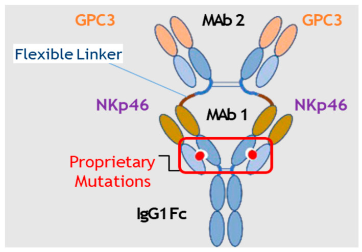
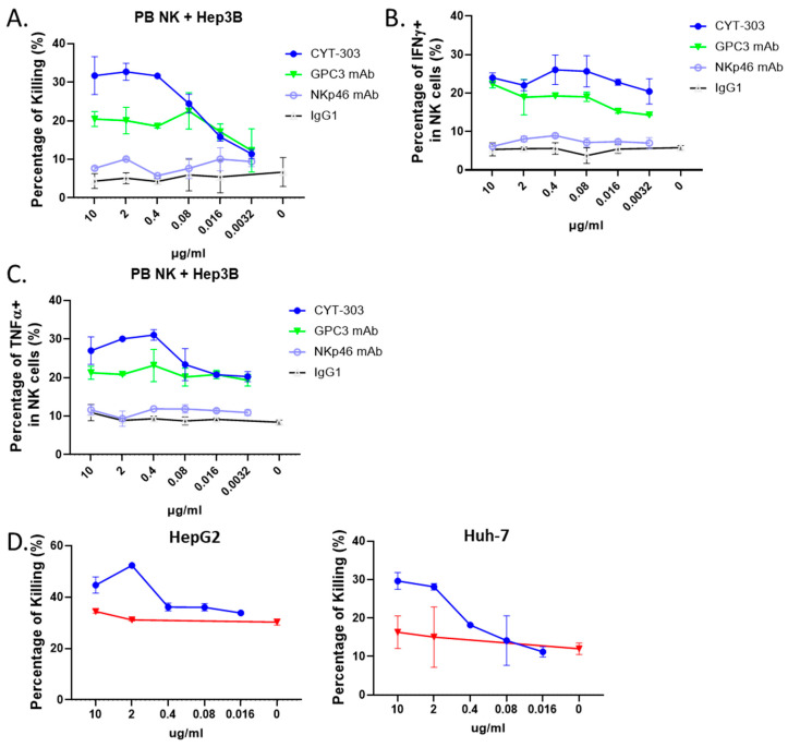
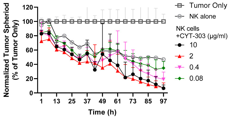
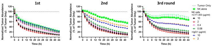
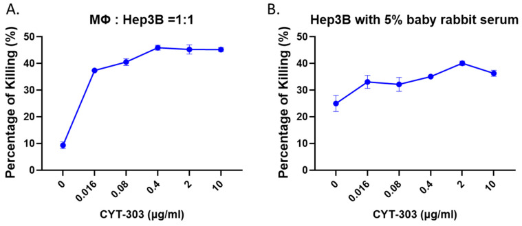
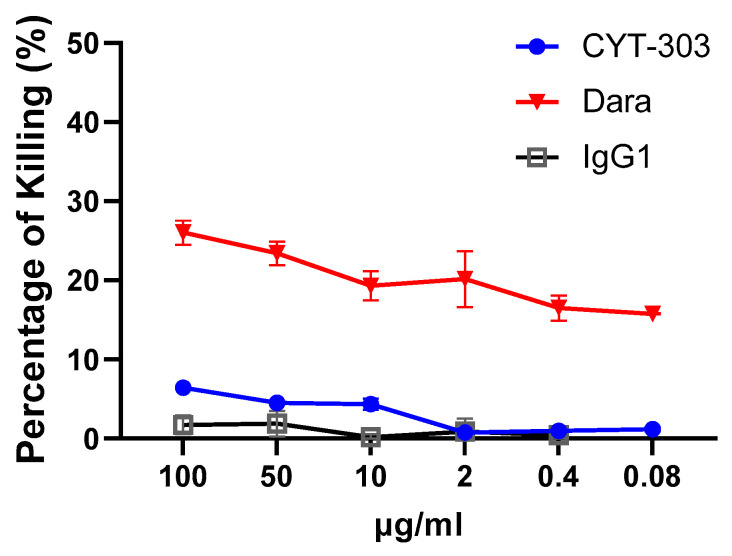
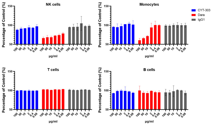
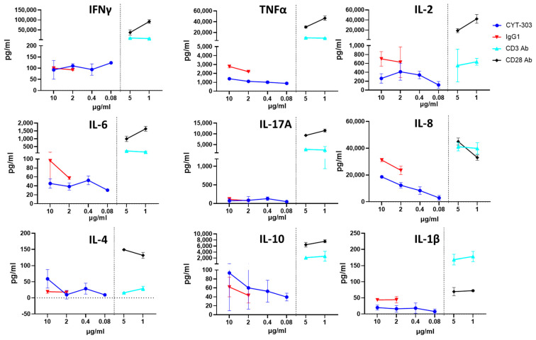
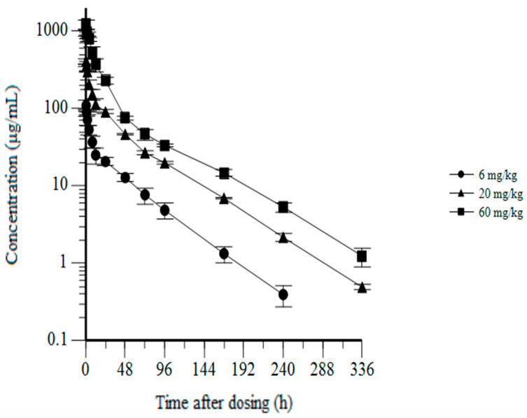
Glypican-3 (GPC3) is an oncofetal antigen that is highly expressed in multiple solid tumors, including hepatocellular carcinoma, and is barely expressed in adult normal tissues except the placenta. NKp46 activation receptor is expressed in all-natural killer (NK) cells, including tumor-infiltrating NK cells. FLEX-NKTM is a platform for the production of tetravalent multifunctional antibody NK cell engagers (NKE). CYT-303 was designed using the FLEX-NK scaffold, incorporating a novel humanized NKp46 binder that does not induce NKp46 internalization and a humanized GPC3 binder that targets the membrane-proximal lobe to mediate NK cell-redirected killing of HCC tumors. CYT-303 shows sub-nanomolar binding affinities to both GPC3 and NKp46. CYT-303 was highly potent and effective in mediating NK cell-redirected cytotoxicity against multiple HCC tumor cell lines and tumor spheroids. More interestingly, it can reverse the dysfunction induced in NK cells following repeated rounds of serial killing of tumors. It also mediated antibody-dependent cellular phagocytosis (ADCP) and complement-dependent cytotoxicity against GPC3-expressing HCC tumors. In vivo, CYT-303 showed no toxicity or cytokine release in cynomolgus monkeys up to the highest dose (60 mg/kg), administered weekly by intravenous infusion for 28 days. These results demonstrate the potential of CYT-303 to be a safe and effective therapy against HCC.

Keywords: GPC3; NK engager; NKp46; bispecific antibody; hepatocellular carcinoma; natural killer cells.


Conflict of interest statement

Antonio Arulanandam, Liang Lin, Hao-Ming Chang, Peng Gao, Armin Rath, Jean Kadouche, Daniel Teper, and Wei Li are employees and hold stock or stock options of Cytovia. Ofer Mandelboim and Armand Bensussan are consultants of Cytovia and hold stock or stock options in Cytovia.

Figures

Figure 1
Figure 1
Diagram of the structure of CYT-303.
Figure 2
Figure 2
Characterization of the anti-NKp46 mAb 09. (A) FACS staining of anti-NKp46 mAbs (9E2 and 09) binding to BW parental cells versus BW transfected cells expressing NKp46 (black and red histograms, respectively). Binding was detected using an anti-mouse IgG AF647 labeled antibody. The filled gray histogram represents staining with secondary antibodies only of the BW parental cells. The background (BG) of BW NKp46 transfectants was similar and is not shown in the figure. The figure shows one representative experiment out of the six performed. (B) FACS staining of anti-NKp46 mAbs (9E2 and 09) on primary activated bulk human NK cells (black histogram). The filled gray histogram represents the staining of NK cells with secondary antibodies only. The figure shows one representative experiment out of the six performed. (C) Human NKp46-Ig was pre-incubated either alone (black histogram) or with anti-NKp46 mAbs (9E2 and 09, red histograms) at 4 °C, followed by FACS staining of BJAB, MCF7, and C1R cells detected with an anti-human IgG PE-labeled antibody. The filled gray histogram represents the staining of cells with secondary antibodies only. The figure shows one representative experiment out of the two performed. (D) Activated bulk NK cell cultures were incubated with the indicated anti-NKp46 mAbs (9E2 and 09) at 4 °C (black histogram) or 37 °C (red histogram) for 8 h, followed by FACS staining with an AF647-labeled anti-mouse secondary antibody. The filled gray histogram represents staining with secondary antibodies only of cells treated at 4 °C. The background of cells treated at 37 °C was similar and is not shown in the figure. The figure shows one representative experiment out of the five performed.
Figure 3
Figure 3
Biochemistry characterization of CYT-303. An amount of 10 μg CYT-303 mixed without (A) or with a reducing agent (B) in the loading buffer and heated at 70 °C for 10 min. The samples were then loaded onto Bolt™ 4 to 12% Bis-Tris protein gel, electrophoresed until the tracking dye reached the bottom of the gel, and stained with AcquaStain protein gel dye.
Figure 4
Figure 4
Binding of CYT-303 to PBNK cells and HCC tumor cells. (A) Purified PB-NKs or Hep3B tumor cells were incubated with the indicated concentrations of CYT-303 or human IgG1 for 60 min at room temperature (RT), and binding was detected by PE-labeled anti-human IgG by flow cytometry. The graphs are presented as means with standard deviations from two replicates. (B) The expression level of CD16 and NKp46 on the purified PBNK cells was assessed using anti-CD16 and anti-NKp46 mAbs. (C) GPC3 expression on liver cancer cell lines Hep3B, Huh-7, and HepG2 using AF647 labeled anti-GPC3 mAb by flow cytometry.
Figure 5
Figure 5
CYT-303 redirected human PBNK cytotoxicity and induced cytokine production. (A) CYT-303 redirected the cytolysis of Hep3B at the indicated concentration of CYT-303 at a fixed E/T ratio of 1 for 5 h, as assessed by flow cytometery using a cell viability dye; (B,C) CYT-303 induced IFNγ and TNFα production of PBNK cells following incubation with Hep3B cells at an E/T ratio of 1 for 5 h were assessed by intracellular staining of PBNKs with anti-IFNγ or TNFα antibodies, respectively. The graph represents data on PBNKs from two donors. (D) Cytolysis of HepG2 and Huh-7 tumors. CYT-303 (blue line) or control human IgG (red line) redirected PBNK cell cytolysis of HepG2 (left) and Huh-7 (right) tumors were evaluated at a fixed E/T = 1 for 5 h and assessed by flow cytometry as described above. The graphs are presented as means with standard deviations from two replicates.
Figure 6
Figure 6
Hep3B-GFP tumor cells were cultured for 2 days in ultra-low attachment U-bottom plates to form tumor spheroids, and then they were incubated with PBNK cells alone or with CYT-303 at the indicated concentrations for 4 days. The remaining tumor spheroids were counted over a period of 4 days. The graph is normalized to the percentage of the tumor only group at each time point and presented as a mean with standard deviation from 3 replicates.
Figure 7
Figure 7
The serial killing of PBNKs against Hep3B-GFP tumors was evaluated with either NK cells alone or in combination with CYT-303 using a fixed E/T ratio (1:2) at each round of killing. Tumor lysis was monitored by the reduction of GFP-positive tumors using the Incucyte Live Analysis System. The graph is normalized to the percentage of the tumor only group at each time point and presented as a mean with a standard deviation from 3 replicates.
Figure 8
Figure 8
CYT-303 mediates ADCP and CDC in vitro. (A) Macrophages were differentiated from purified monocytes following culture with M-CSF for 5 days. CYT-303-induced macrophage phagocytosis of Hep3B-GFP tumor cells was assessed in a 4-h assay using viability dye staining of tumor cells by flow cytometry. The graph is presented as a mean with a standard deviation from three replicates. (B) Hep3B tumor cells were incubated with CYT-303 and baby rabbit complement for 4 h, and tumor cell lysis was assessed by flow cytometry using cell viability dye. The graphs are presented as means with standard deviations from four replicates.
Figure 9
Figure 9
CYT-303 did not induce NK cell fratricide. NK cell fratricide by CYT-303 was evaluated using purified PBNK in the presence of CYT-303, Daratumumab, or human IgG for 24 h. Fratricide was assessed by staining PBNK cells with a live dead cell dye and analyzed by flow cytometry. The graph represents data on PBNKs from two donors.
Figure 10
Figure 10
CYT-303 did not induce immune cell depletion. Human PBMCs were incubated with different concentrations of CYT-303, Daratumumab, or human IgG1 for 24 h. Depletion of the immune cell subsets was analyzed by flow cytometry using immune cell subset-specific antibodies and compared to a human IgG control. The graph represents data on PBNKs from two donors.
Figure 11
Figure 11
CYT-303 did not induce significant non-specific cytokine release in vitro. Human PBMCs were incubated with indicated concentrations of CYT-303, anti-CD3, or anti-CD28 (TGN1412) mAbs for 48 h. Cytokine release was assessed by a multiplex cytokine immunoassay. The graph represents data on PBNKs from two donors.
Figure 12
Figure 12
CYT-303 single-dose pharmacokinetics in cynomolgus monkeys. Cynomolgus monkeys were dosed with CYT-303 by intravenous infusion at the indicated dose levels. Blood concentrations were measured up to 336 h post-dosing using a PK immunoassay. Three monkeys per group were evaluated.

References

    1. Birch G.C., Fehniger T.F., Romee R. Biology of NK cells and NK cells in clinic. In: Ghobadi A., DiPersio J.F., editors. Gene and Cellular Immunotherapy for Cancer. Springer International Publishing; Cham, Switzerland: 2022. pp. 293–325.
    1. Liu E., Marin D., Banerjee P., Macapinlac H.A., Thompson P., Basar R., Nassif Kerbauy L., Overman B., Thall P., Kaplan M., et al. Use of CAR-Transduced Natural Killer Cells in CD19-Positive Lymphoid Tumors. N. Engl. J. Med. 2020;382:545–553. doi: 10.1056/NEJMoa1910607. - DOI - PMC - PubMed
    1. Gazit R., Gruda R., Elboim M., Arnon T.I., Katz G., Achdout H., Hanna J., Qimron U., Landau G., Greenbaum E., et al. Lethal influenza infection in the absence of the natural killer cell receptor gene Ncr1. Nat. Immunol. 2006;7:517–523. doi: 10.1038/ni1322. - DOI - PubMed
    1. Sivori S., Vitale M., Morelli L., Sanseverino L., Augugliaro R., Bottino C., Moretta L., Moretta A. p46, a novel natural killer cell-specific surface molecule that mediates cell activation. J. Exp. Med. 1997;186:1129–1136. doi: 10.1084/jem.186.7.1129. - DOI - PMC - PubMed
    1. Gauthier L., Morel A., Anceriz N., Rossi B., Blanchard-Alvarez A., Grondin G., Trichard S., Cesari C., Sapet M., Bosco F., et al. Multifunctional Natural Killer Cell Engagers Targeting NKp46 Trigger Protective Tumor Immunity. Cell. 2019;177:1701–1713.e16. doi: 10.1016/j.cell.2019.04.041. - DOI - PubMed

Publication types