

Cancer Spheroids and Organoids as Novel Tools for Research and Therapy: State of the Art and Challenges to Guide Precision Medicine
- PMID: 37048073
- PMCID: PMC10093533
- DOI: 10.3390/cells12071001
Cancer Spheroids and Organoids as Novel Tools for Research and Therapy: State of the Art and Challenges to Guide Precision Medicine
Abstract
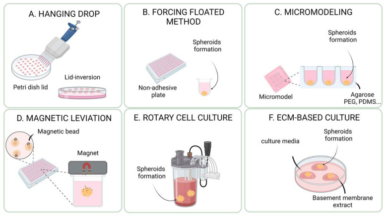
Spheroids and organoids are important novel players in medical and life science research. They are gradually replacing two-dimensional (2D) cell cultures. Indeed, three-dimensional (3D) cultures are closer to the in vivo reality and open promising perspectives for academic research, drug screening, and personalized medicine. A large variety of cells and tissues, including tumor cells, can be the starting material for the generation of 3D cultures, including primary tissues, stem cells, or cell lines. A panoply of methods has been developed to generate 3D structures, including spontaneous or forced cell aggregation, air-liquid interface conditions, low cell attachment supports, magnetic levitation, and scaffold-based technologies. The choice of the most appropriate method depends on (i) the origin of the tissue, (ii) the presence or absence of a disease, and (iii) the intended application. This review summarizes methods and approaches for the generation of cancer spheroids and organoids, including their advantages and limitations. We also highlight some of the challenges and unresolved issues in the field of cancer spheroids and organoids, and discuss possible therapeutic applications.
Keywords: 3D cell culture; cell and gene therapy; drug screening; immunotherapy; personalized medicine; tissue engineering.
Conflict of interest statement
The authors declare no conflict of interest.
Figures


Similar articles
-
Historical evolution of spheroids and organoids, and possibilities of use in life sciences and medicine.Biotechnol J. 2021 May;16(5):e2000463. doi: 10.1002/biot.202000463. Epub 2021 Jan 25. Biotechnol J. 2021. PMID: 33491924 Review.
-
[Spheroids to organoids: Solid cancer models for anticancer drug discovery].Bull Cancer. 2022 Jan;109(1):49-57. doi: 10.1016/j.bulcan.2021.09.019. Epub 2021 Nov 27. Bull Cancer. 2022. PMID: 34848046 Review. French.
-
Modeling neoplastic disease with spheroids and organoids.J Hematol Oncol. 2020 Jul 16;13(1):97. doi: 10.1186/s13045-020-00931-0. J Hematol Oncol. 2020. PMID: 32677979 Free PMC article. Review.
-
Patient-Derived Organoids: A Game-Changer in Personalized Cancer Medicine.Stem Cell Rev Rep. 2025 Jan;21(1):211-225. doi: 10.1007/s12015-024-10805-4. Epub 2024 Oct 21. Stem Cell Rev Rep. 2025. PMID: 39432173 Review.
-
Generation of Three-Dimensional Spheroids/Organoids from Two-Dimensional Cell Cultures Using a Novel Stamp Device.J Vis Exp. 2025 Mar 28;(217). doi: 10.3791/67787. J Vis Exp. 2025. PMID: 40227993
Cited by
-
Omics-based molecular classifications empowering in precision oncology.Cell Oncol (Dordr). 2024 Jun;47(3):759-777. doi: 10.1007/s13402-023-00912-8. Epub 2024 Jan 31. Cell Oncol (Dordr). 2024. PMID: 38294647 Review.
-
Deep Learning for Predicting Spheroid Viability: Novel Convolutional Neural Network Model for Automating Quality Control for Three-Dimensional Bioprinting.Bioengineering (Basel). 2025 Jan 1;12(1):28. doi: 10.3390/bioengineering12010028. Bioengineering (Basel). 2025. PMID: 39851302 Free PMC article.
-
Effect of Neutron Radiation on 10BPA-Loaded Melanoma Spheroids and Melanocytes.Cells. 2025 Feb 6;14(3):232. doi: 10.3390/cells14030232. Cells. 2025. PMID: 39937023 Free PMC article.
-
State of the Art in 3D Culture Models Applied to Thyroid Cancer.Medicina (Kaunas). 2024 Mar 22;60(4):520. doi: 10.3390/medicina60040520. Medicina (Kaunas). 2024. PMID: 38674166 Free PMC article. Review.
-
GRHL2-HER3 and E-cadherin mediate EGFR-bypass drug resistance in lung cancer cells.Front Cell Dev Biol. 2025 Jan 17;12:1511190. doi: 10.3389/fcell.2024.1511190. eCollection 2024. Front Cell Dev Biol. 2025. PMID: 39897079 Free PMC article.
References
-
- Weeber F., van de Wetering M., Hoogstraat M., Dijkstra K.K., Krijgsman O., Kuilman T., Gadellaa-van Hooijdonk C.G.M., van der Velden D.L., Peeper D.S., Cuppen E.P.J.G., et al. Preserved genetic diversity in organoids cultured from biopsies of human colorectal cancer metastases. Proc. Natl. Acad. Sci. USA. 2015;112:13308–13311. doi: 10.1073/pnas.1516689112. - DOI - PMC - PubMed
-
- Schutte M., Risch T., Abdavi-Azar N., Boehnke K., Schumacher D., Keil M., Yildiriman R., Jandrasits C., Borodina T., Amstislavskiy V., et al. Molecular dissection of colorectal cancer in pre-clinical models identifies biomarkers predicting sensitivity to EGFR inhibitors. Nat. Commun. 2017;8:14262. doi: 10.1038/ncomms14262. - DOI - PMC - PubMed
Publication types
MeSH terms
LinkOut - more resources
Full Text Sources
Medical
