Review
. 2023 Mar 25;12(7):1008.
doi: 10.3390/cells12071008.

A Review of the Biosynthesis and Structural Implications of Insulin Gene Mutations Linked to Human Disease

Affiliations
Review

A Review of the Biosynthesis and Structural Implications of Insulin Gene Mutations Linked to Human Disease

Sara Ataie-Ashtiani et al. Cells. .

Abstract

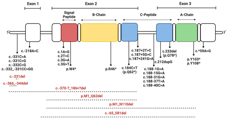
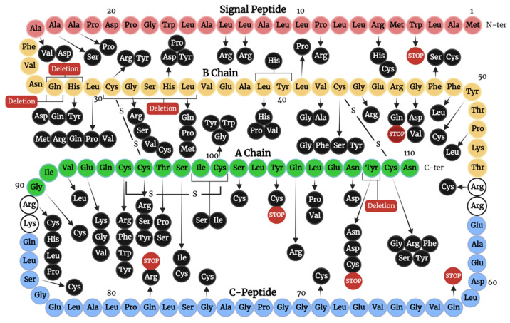
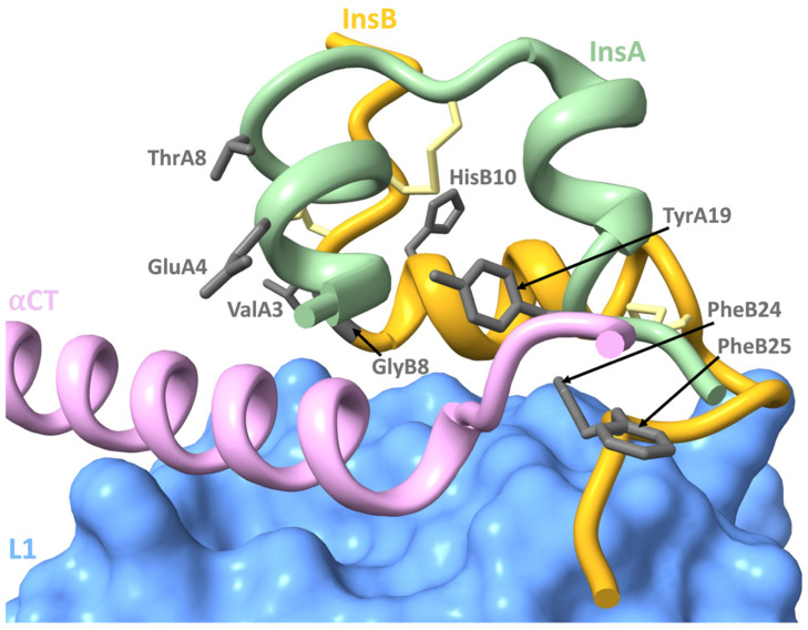
The discovery of the insulin hormone over 100 years ago, and its subsequent therapeutic application, marked a key landmark in the history of medicine and medical research. The many roles insulin plays in cell metabolism and growth have been revealed by extensive investigations into the structure and function of insulin, the insulin tyrosine kinase receptor (IR), as well as the signalling cascades, which occur upon insulin binding to the IR. In this review, the insulin gene mutations identified as causing disease and the structural implications of these mutations will be discussed. Over 100 studies were evaluated by one reviewing author, and over 70 insulin gene mutations were identified. Mutations may impair insulin gene transcription and translation, preproinsulin trafficking and proinsulin sorting, or insulin-IR interactions. A better understanding of insulin gene mutations and the resultant pathophysiology can give essential insight into the molecular mechanisms underlying impaired insulin biosynthesis and insulin-IR interaction.

Keywords: insulin biosynthesis; insulin gene mutations; mutant insulin; neonatal diabetes.


Conflict of interest statement

The authors declare no conflict of interest.

Figures

Figure 1
Figure 1
Schematic of the stages of insulin biosynthesis. 1. Insulin gene containing three exons separated by two introns. 2. Preproinsulin produced via the transcription and translation of the insulin gene. 3. Proinsulin produced from preproinsulin after the signal peptide is cleaved off by signal peptidase. 4. Mature insulin produced as the C-peptide is cleaved from proinsulin within secretory vesicles. Displayed domains: the amino-terminal (N-ter) signal peptide (red), the B-chain (yellow), the C-peptide (blue), and the carboxy-terminal (C-ter) A-chain (green).
Figure 2
Figure 2
Schematic of the life cycle of insulin biosynthesis. After transcription within the nucleus (step 1), translation occurs at the cytosolic face of the Endoplasmic Reticulum (ER) (step 2), producing preproinsulin. Within the ER, the signal peptide is cleaved, disulfide bonds form, and the peptide is folded. Proinsulin moves into the Golgi body where it is sorted and packed into secretory vesicles (step 3). Within these vesicles, the C-peptide is cleaved off by prohormone convertases (PC1/3 and PC2) and carboxypeptidase E (CPE), forming mature insulin (step 4). Insulin is secreted in response to secretory signals that result in membrane depolarisation and calcium influx into the cell (top left). Insulin binds to IR (step 5) and the resultant receptor activation initiates intracellular signalling cascades.
Figure 3
Figure 3
Schematic of the location of human insulin gene mutations affecting transcription and translation. Deletions are shown in red text and using dashed lines. Throughout this review, two naming systems are used. For mutations within the insulin gene, the “coding” system, as indicated by the presence of c. in front of the mutation name, is used. For mutations that affect the protein sequence, the “protein” naming system is used, as indicated by the presence of p. in front of the mutation name. Mutations resulting in protein truncations are annotated using the protein naming system, followed by “*”. Mutations starting with “–“ are located in the promoter or before the start of the coding sequence of the gene, while those starting with “*” are located after the coding sequence of the gene.
Figure 4
Figure 4
Schematic demonstrating the amino acid sequence of human preproinsulin with annotations of the location of insulin mutations impacting trafficking, processing, and IR interactions. Displayed domains: the amino-terminal (N-ter) signal peptide (red), B-chain (yellow), C-peptide (blue), and the carboxy-terminal (C-ter) A-chain (green). White residues are those which are cleaved during the conversion of proinsulin to insulin. Mutations resulting in stop codons and deletions are seen in red.
Figure 5
Figure 5
Schematic of wild-type insulin interacting with the insulin receptor (IR). Side chains of insulin residues key for IR interaction, with identified mutations, displayed in grey. Displayed domains: insulin B-chain (gold), insulin A-chain (light green), IR L1 (blue), IR αCT’ (pink). Insulin disulfide bonds are seen in yellow.

References

    1. White M.F., Kahn C.R. Insulin action at a molecular level—100 years of progress. Mol. Metab. 2021;52:101304. doi: 10.1016/j.molmet.2021.101304. - DOI - PMC - PubMed
    1. Boucher J., Kleinridders A., Kahn C.R. Insulin receptor signaling in normal and insulin-resistant states. Cold Spring Harb. Perspect. Biol. 2014;6:a009191. doi: 10.1101/cshperspect.a009191. - DOI - PMC - PubMed
    1. Hatting M., Tavares C.D.J., Sharabi K., Rines A.K., Puigserver P. Insulin regulation of gluconeogenesis. Ann. N. Y. Acad. Sci. 2018;1411:21–35. doi: 10.1111/nyas.13435. - DOI - PMC - PubMed
    1. Støy J., De Franco E., Ye H., Park S.-Y., Bell G.I., Hattersley A.T. In celebration of a century with insulin—Update of insulin gene mutations in diabetes. Mol. Metab. 2021;52:101280. doi: 10.1016/j.molmet.2021.101280. - DOI - PMC - PubMed
    1. Misra S., Owen K.R. Genetics of Monogenic Diabetes: Present Clinical Challenges. Curr. Diabetes Rep. 2018;18:141. doi: 10.1007/s11892-018-1111-4. - DOI - PMC - PubMed