Review
. 2023 Mar 25;12(7):1012.
doi: 10.3390/cells12071012.

Microglia Mediated Neuroinflammation in Parkinson's Disease

Affiliations
Review

Microglia Mediated Neuroinflammation in Parkinson's Disease

Sevim Isik et al. Cells. .

Abstract

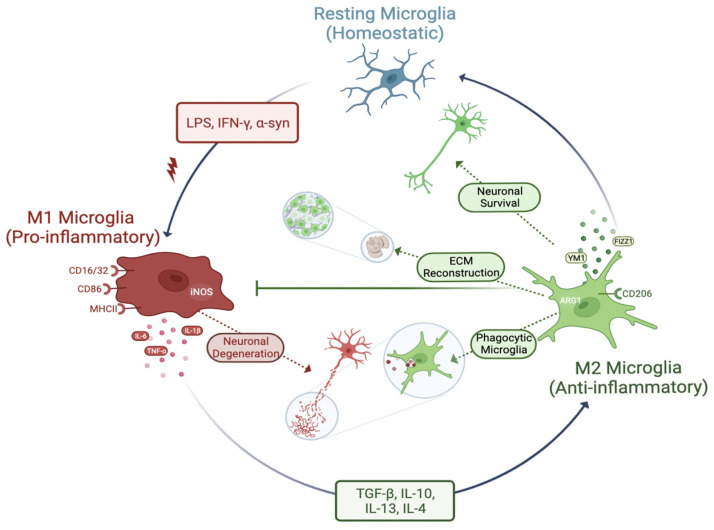
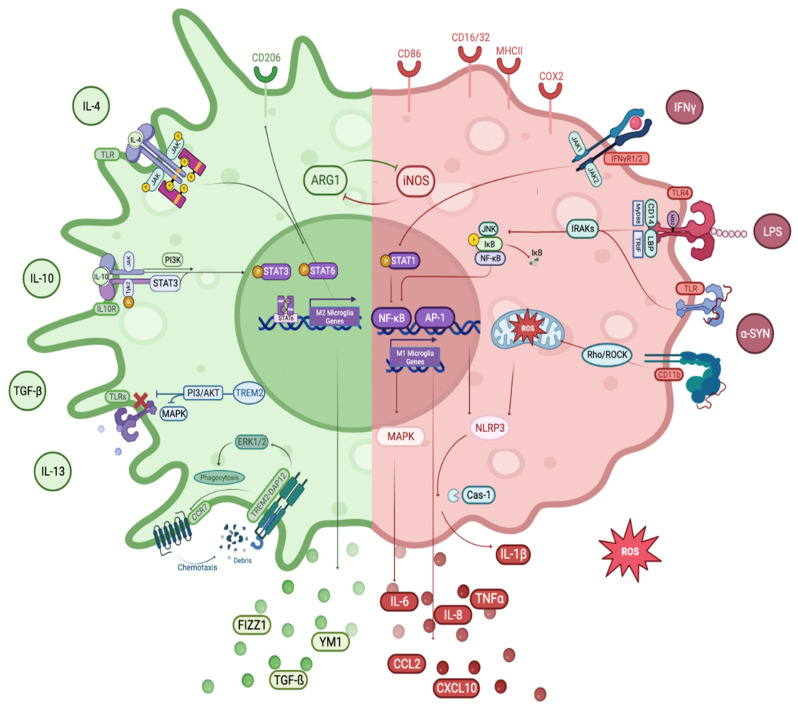
Parkinson's Disease (PD) is the second most common neurodegenerative disorder seen, especially in the elderly. Tremor, shaking, movement problems, and difficulty with balance and coordination are among the hallmarks, and dopaminergic neuronal loss in substantia nigra pars compacta of the brain and aggregation of intracellular protein α-synuclein are the pathological characterizations. Neuroinflammation has emerged as an involving mechanism at the initiation and development of PD. It is a complex network of interactions comprising immune and non-immune cells in addition to mediators of the immune response. Microglia, the resident macrophages in the CNS, take on the leading role in regulating neuroinflammation and maintaining homeostasis. Under normal physiological conditions, they exist as "homeostatic" but upon pathological stimuli, they switch to the "reactive state". Pro-inflammatory (M1) and anti-inflammatory (M2) phenotypes are used to classify microglial activity with each phenotype having its own markers and released mediators. When M1 microglia are persistent, they will contribute to various inflammatory diseases, including neurodegenerative diseases, such as PD. In this review, we focus on the role of microglia mediated neuroinflammation in PD and also signaling pathways, receptors, and mediators involved in the process, presenting the studies that associate microglia-mediated inflammation with PD. A better understanding of this complex network and interactions is important in seeking new therapies for PD and possibly other neurodegenerative diseases.

Keywords: M1 phenotype; M2 phenotype; Parkinson’s Disease; anti-inflammatory phenotype; microglial activation; neuroinflammation; α-synuclein.


Conflict of interest statement

The authors declare no conflict of interest.

Figures

Figure 1
Figure 1
M1 and M2 microglia phenotypes. Under physiological circumstances, microglia exhibit the homeostatic microglia phenotype. Depending on the environment in which they are reactive and the factors in which they are stimulated, they can change into “pro-inflammatory (M1)” or “anti-inflammatory (M2)” phenotypes. Microglia play important roles in maintaining the health of neurons, including pruning and remodeling synapses, controlling myelination, and removing pathological proteins that are misfolded through neurogenesis and phagocytosis. Microglia are also responsible for maintaining the homeostasis of brain tissue. Additionally, depending on the type of activation, microglia secrete numerous trophic factors, cytokines, and chemokines to aid in neuronal survival. Pathogenic molecules, such as LPS and/or IFN, or protein aggregates, such as α-syn, stimulate microglia into the pro-inflammatory phenotype, which then releases inflammatory molecules, such as ROS and other pro-inflammatory cytokines, including IL-1β, iNOS, TNFα, and others. Persistent exposure of microglia to these inflammatory mediators may result in neuronal damage. Contrarily, mediators, such as TGF-β, IL-4, IL-10, and IL-13, induce the M1 to M2 transition. The M2 phenotype of microglia contribute to the processes in phagocytosis, ECM rebuilding, and neuronal survival by secreting such factors as Ym1 and FIZZ1.
Figure 2
Figure 2
Overview of microglial M1 and M2 signaling pathways in PD. The right side of the figure represents the M1 microglial phenotype and its related signaling pathways. LPS, which is one of the main M1 microglia activators, binds to MD2-bound TLR4 on the cell surface via LBP (LPS-binding protein) and CD14, which acts as a co-receptor. The resulting complex binds to TRIF and Myd88 by interacting with the cytoplasmic domain of TLRs and individual TIR domains. Activated TLR phosphorylates the IKK complex, which consists of MAP kinases, such as IKKβ, JNK, through autophosphorylation of IRAKs, thereby inducing translocation and activation of transcription factors NF-κB and AP-1 and initiates upregulation of M1-associated gene transcription. M1 activation by IFNγ occurs by initiation of the signaling pathway by IFNγR1/2, phosphorylation of STAT1 and interferon regulatory factors, and translocation to the nucleus via JAK1/2. Aggregates of α-syn released into the extracellular space bind to TLR2 following the working mechanism of TLR2s, triggering activation of NF-kB and subsequent NF-kB-dependent upregulation of NLRP3 and production of proinflammatory cytokines. NLRP3 activation provides caspase-1-mediated release of IL-1ß and IL-8. In addition, α-syn clusters are recognized by CD11b and induce mitochondrial ROS generation by impairing mitochondrial function via the Rho/ROCK pathway. In addition, the activation of the M1 phenotype contributes to the regulation of intracellular iNOS, cell surface markers (CD86, CD16/32, and MHC II), M1-related pro-inflammatory cytokines (IL-1β, IL-6, IL-12, IL-17, IL-18, IL-23, and TNFα), and chemokines (CCL2, CXCL10). The left side of the figure represents the M2 microglial phenotype and its related signaling pathways. M2 status is mainly induced by anti-inflammatory stimuli, such as IL-4, IL-10, IL-13, TGF-β, and glucocorticoids. IL-4 binds to IL-4R causing phosphorylation of JAKs/STAT6 and translocation of STAT6 to the nucleus. Activated STAT6 specifically leads to the transcription of M2-related genes, including intracellular components, such as CD206 and cytokine signal suppressor 3 (SOCS3). IL-10 binds to IL-10R1/2, enabling STAT3 to be phosphorylated and translocated to the nucleus via the JAK/STAT signal cascade and PI3K. STAT3 translocation inhibits M1-related proinflammatory cytokines and upregulation of IL-10 and TGF-β. TGF-β increases ARG1 expression and decreases iNOS and COX2. M2 microglia activation releases anti-inflammatory molecules, such as Arg-1, IGF-1, Ym1, and FIZZ1, which contribute to matrix deposition and wound healing. With the increase of the M2 phenotype, TREM2 inhibits the PI3/AKT signaling pathway, TLRs, and MAPK and provides the transition to the M2 phenotype and the inhibition of the M1 phenotype. In addition, the TREM2/DAP12 complex stimulates ERK1/2 by regulating actin polymerization and cytoskeleton with ERK1/2 activation. This activation increases the expression of CCR7 on the cell surface and provides chemotactic migration towards CCR7 ligands. In addition, TREM2 activation stimulates microglial phagocytosis in the same way. Activation of the PI3K/Akt pathway by TREM2/DAP12 contributes to the regulation of NF-κB and inhibition of TLR signaling by blocking MAPK signaling.

Similar articles

Cited by

References

    1. Kalia L.V., Lang A.E. Parkinson’s Disease. Lancet. 2015;386:896–912. doi: 10.1016/S0140-6736(14)61393-3. - DOI - PubMed
    1. Tanner C.M., Goldman S.M. Epidemiology of Parkinson’s Disease. Neurol. Clin. 1996;14:317–335. doi: 10.1016/S0733-8619(05)70259-0. - DOI - PMC - PubMed
    1. Wang W., Song N., Jia F., Tang T., Bao W., Zuo C., Xie J., Jiang H. Genomic DNA Levels of Mutant Alpha-synuclein Correlate with Non-Motor Symptoms in an A53T Parkinson’s Disease Mouse Model. Neurochem. Int. 2018;114:71–79. doi: 10.1016/j.neuint.2018.01.006. - DOI - PubMed
    1. Frost J.J., Rosier A.J., Reich S.G., Smith J.S., Ehlers M.D., Snyder S.H., Ravert H.T., Dannals R.F. Positron Emission Tomographic Imaging of the Dopamine Transporter with 11C-WIN 35,428 Reveals Marked Declines in Mild Parkinson’s Disease. Ann. Neurol. 1993;34:423–431. doi: 10.1002/ana.410340331. - DOI - PubMed
    1. Todd K.L., Lipski J., Freestone P.S. Subthalamic Nucleus Exclusively Evokes Dopamine Release in the Tail of the Striatum. J. Neurochem. 2022;162:417–429. doi: 10.1111/jnc.15677. - DOI - PMC - PubMed

Publication types

MeSH terms