. 2023 Mar 30;12(7):1046.
doi: 10.3390/cells12071046.

Loss-of-Function Variants in DRD1 in Infantile Parkinsonism-Dystonia

Affiliations

Loss-of-Function Variants in DRD1 in Infantile Parkinsonism-Dystonia

Kimberley M Reid et al. Cells. .

Abstract

The human dopaminergic system is vital for a broad range of neurological processes, including the control of voluntary movement. Here we report a proband presenting with clinical features of dopamine deficiency: severe infantile parkinsonism-dystonia, characterised by frequent oculogyric crises, dysautonomia and global neurodevelopmental impairment. CSF neurotransmitter analysis was unexpectedly normal. Triome whole-genome sequencing revealed a homozygous variant (c.110C>A, (p.T37K)) in DRD1, encoding the most abundant dopamine receptor (D1) in the central nervous system, most highly expressed in the striatum. This variant was absent from gnomAD, with a CADD score of 27.5. Using an in vitro heterologous expression system, we determined that DRD1-T37K results in loss of protein function. Structure-function modelling studies predicted reduced substrate binding, which was confirmed in vitro. Exposure of mutant protein to the selective D1 agonist Chloro APB resulted in significantly reduced cyclic AMP levels. Numerous D1 agonists failed to rescue the cellular defect, reflected clinically in the patient, who had no benefit from dopaminergic therapy. Our study identifies DRD1 as a new disease-associated gene, suggesting a crucial role for the D1 receptor in motor control.

Keywords: DRD1; dopamine; dystonia; parkinsonism.


Conflict of interest statement

M.A.K. is a co-founder of Bloomsbury Therapeutics.

Figures

Figure 1
Figure 1
Schematic representation of direct and indirect pathways of the basal ganglia. Left: The direct and indirect pathways regulate voluntary movement and form a key part of the cortico-basal ganglia-thalamic network. The striatum receives inputs including dopaminergic fibres from the substantia nigra, as well as GABAergic (inhibitory) inputs from the globus pallidus pars externa and glutamatergic (excitatory) inputs from the cortex, thalamus and subthalamic nucleus. In turn, it provides inhibitory output via GABergic medium spiny neurons (MSNs) to the globus pallidus pars interna and externa and the substantia nigra. The balance of these outputs determines the relative activity of the direct (prokinetic) and indirect (antikinetic) pathways by modulating thalamic excitatory output to the motor cortex. Right: Dopamine in the striatum has opposite effects depending on whether the postsynaptic MSN expresses D1 or D2 receptors, exciting the former—which contribute to the direct pathway—and inhibiting the latter—which contribute to the indirect pathway.
Figure 2
Figure 2
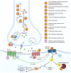
Dopamine pathway in the pre-synaptic dopaminergic neuron: key enzymes, proteins and associated diseases. Schematic representation of a dopaminergic synapse. Dopamine is synthesised from tyrosine in the presynaptic cell: tyrosine is converted to L-dopa by tyrosine hydroxylase, with tetrahydrobiopterin as a cofactor, then L-dopa is decarboxylated to dopamine by aromatic l-amino acid decarboxylase (AADC). Dopamine is packaged into presynaptic vesicles by the vesicular monoamine transporter 2 (VMAT2), before being released into the synapse. On the post-synaptic membrane, dopamine binds D1-like and D2-like receptors, causing the activation or inhibition of adenylyl cyclase, leading to increase or decrease of intracellular cAMP and downstream signalling pathways mediated by protein kinase A. Stars indicate genetic diseases associated with defects in these pathways.
Figure 3
Figure 3
Clinical features and genetic findings in proband. (A) Family pedigree, with affected individuals indicated by black shading. (BD) Images of proband aged approximately four years depicting phenotype. (B) Bilateral fixed upwards eye deviation during an oculogyric crisis. (C) Dystonic posturing of trunk and limbs; note striatal toe on the right. (D) Opisthotonic posturing and dystonic jaw opening. (E) Alignment of cross-species DRD1 protein sequences in human, chimp, macaque, rat, mouse, platypus, chicken, frog and zebrafish showing conservation of the amino acid and surrounding region. Black box indicates relevant amino acid. (F) Sanger sequencing analysis confirming the DRD1 variant. Sequence chromatograms show heterozygous variants at position c.110 in both parents, and a homozygous variant in proband.
Figure 4
Figure 4
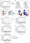
Effect of T37K variant on cAMP production and ligand binding. (A) HEK-293T cells transfected with either DRD1-WT or DRD1-T37K were treated with increasing concentrations of Chloro APB for 10 min and cAMP levels recorded. Graph displays the mean inverted luminescence +/− SEM (n = 6). ns = not significant, * p < 0.05, ** p < 0.01, *** p < 0.005, **** p < 0.001 (2-way ANOVA between DRD1-WT and DRD1-T37K). (BG) Structures of DRD1 wildtype and modelled T37K mutant proteins. The side view of the DRD1 proteins showing the angles between the helices TM1 and TM2 with the wildtype protein (PDB ID: 7JVP) in pink (B) and T37K mutant in blue (C). The orientation of T37 (D) and K37 (E) in the DRD1 wildtype (pink) and T37K mutant proteins (blue). The amino acids are represented in single letter codes and coloured by atom-type. Coulombic potential-based surface rendering of the wildtype (F) and the T37K mutant (G) proteins around the sites depicted in (C,D). Red, blue and white correspond to negative, positive and no charge on the residues, respectively. The graphical representation of the protein structures was created using the molecular graphics program Chimera (http://www.cgl.ucsf.edu/chimera/ accessed on 1 December 2022). (H) DA binding to either D1-WT or D1-T37K was investigated using increasing concentrations of (3H) DA. Data are presented as the mean +/− SEM relative to untransfected treated cells (2-way ANOVA, n = 6, p < 0.05). (IL) Transfected HEK-293T cells treated with clinically relevant D1 agonists (I) pramipexole, (J) apomorphine, (K) rotigotine and (L) bromocriptine. Graph displays the mean inverted luminescence +/- SEM (n = 6). ns = not significant, * p < 0.05, ** p < 0.01, *** p < 0.005, **** p < 0.001 (2-way ANOVA between D1-WT and D1-T37K).

References

    1. Klein M.O., Battagello D.S., Cardoso A.R., Hauser D.N., Bittencourt J.C., Correa R.G. Dopamine: Functions, Signaling, and Association with Neurological Diseases. Cell. Mol. Neurobiol. 2019;39:31–59. doi: 10.1007/s10571-018-0632-3. - DOI - PMC - PubMed
    1. Lee H.J., Weitz A.J., Bernal-Casas D., Duffy B.A., Choy M.K., Kravitz A.V., Kreitzer A.C., Lee J.H. Activation of Direct and Indirect Pathway Medium Spiny Neurons Drives Distinct Brain-wide Responses. Neuron. 2016;91:412–424. doi: 10.1016/j.neuron.2016.06.010. - DOI - PMC - PubMed
    1. Jackson D.M., Westlind-Danielsson A. Dopamine receptors: Molecular biology, biochemistry and behavioural aspects. Pharmacol. Ther. 1994;64:291–370. doi: 10.1016/0163-7258(94)90041-8. - DOI - PubMed
    1. Lobo M.K. Past, Present, and Future. 1st ed. Volume 89. Elsevier Inc.; Amsterdam, The Netherlands: 2009. Molecular Profiling of Striatonigral and Striatopallidal Medium Spiny Neurons; pp. 1–35. International Review of Neurobiology. - DOI - PubMed
    1. Kravitz A.V., Freeze B.S., Parker P.R.L., Kay K., Thwin M.T., Deisseroth K., Kreitzer A.C. Regulation of parkinsonian motor behaviours by optogenetic control of basal ganglia circuitry. Nature. 2010;466:622–626. doi: 10.1038/nature09159. - DOI - PMC - PubMed

Publication types

Supplementary concepts