Review
. 2023 Mar 30;12(7):1057.
doi: 10.3390/cells12071057.

Cracking the Code of Neuronal Cell Fate

Affiliations
Review

Cracking the Code of Neuronal Cell Fate

Giovanna Morello et al. Cells. .

Abstract

Transcriptional regulation is fundamental to most biological processes and reverse-engineering programs can be used to decipher the underlying programs. In this review, we describe how genomics is offering a systems biology-based perspective of the intricate and temporally coordinated transcriptional programs that control neuronal apoptosis and survival. In addition to providing a new standpoint in human pathology focused on the regulatory program, cracking the code of neuronal cell fate may offer innovative therapeutic approaches focused on downstream targets and regulatory networks. Similar to computers, where faults often arise from a software bug, neuronal fate may critically depend on its transcription program. Thus, cracking the code of neuronal life or death may help finding a patch for neurodegeneration and cancer.

Keywords: apoptosis; drug repurposing; drug targets; functional enrichment; neurological disease; neurotrophic factors; regulatory network; survival; transcriptional analysis.


Conflict of interest statement

The authors declare no conflict of interest.

Figures
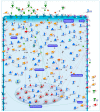
Figure 1
Figure 1
Transcriptional Profiles of Cell Fate Transitions Reveal Early Drivers of Neuronal Apoptosis and Survival. In our recent work [43], we identified a core set of 175 genes exhibiting a significant opposite expression trend during the early commitment phase (0.5 h, 1 h and 3 h) of CGNs apoptosis or its rescue by three NFs (Pacap, Igf1 and SP). The illustrative map shows the biological function and sub-cellular localization of the encoded proteins of core set genes. Significant gene expression changes are shown with “thermometer-like” figures. Numbers indicate time points: ① 0.5 h, ② 1 h, and ③ 3 h following induction of CGNs apoptosis and rescue by NFs. For each time-point, the upward thermometers (red) indicate gene transcripts up-regulated by NFs treatment, while downward thermometers (blue) indicate genes down-regulated. The pathway map was created using MetaCore Pathway Map Creator tool (GeneGo). Further explanations are provided at https://portal.genego.com/legends/MetaCoreQuickReferenceGuide.pdf. (accessed on 14 February 2023).
Figure 2
Figure 2
Core set genes are involved in the etio-pathogenesis of multiple cancer and neurological disorders. (A) Pie chart representation of core set genes implicated in human diseases. Disease enrichment analysis was performed with DAVID bioinformatics resources, including OMIM, KEGG DISEASE, and GAD catalogs. (B) The protein–protein interaction network of the 29 core set genes previously associated with cognitive/mental diseases. The network was built using the STRING website and visualized by Cytoscape (version: 3.8.2), by mapping the ‘degree parameter’ to node size. As the node size increased, the value of the connectivity degree of node genes increased. Differently colored ‘edges’ indicate the type of evidence supporting each interaction: dark purple: co-expression; light purple: physical interaction; light blue: co-localization; light green: shared protein domain; orange: predicted; grey: other. (C) Histogram of the most significantly enriched transcriptional signatures from iLINCS positively correlated with apoptotic CGN-related expression changes of core set genes. The significance of each disease related signature is represented by the enrichment scores value (−log10 (p-value)). For more details, please refer to the original work [43].
Figure 3
Figure 3
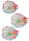
Reverse-engineering of transcriptional changes identifies key transcription factors at the intersection of neuronal apoptosis and survival. Inferring transcriptional gene regulatory networks of core set genes identify nine transcription factors (Hoxd9, Maf, Nr4a1, Cebpb, Olig2, Onecut2, Spdef, Twist2, Nfyb), which may act as upstream regulators of neuronal cell fate [43]. Transcription regulatory network analysis generated at each time point (0.5 h, 1 h, 3 h) following induction of apoptosis or rescue by NFs emphasizes how temporally distinct apoptosis and survival-inducing signals are orchestrated by the action of interconnected and temporally ordered TFs. Regulatory networks are visualized by Cytoscape and for each time-point the node color is consistent with the expression logFC of each gene: genes in blue are down-regulated by NFs treatment, while genes in red are up-regulated. Transcription factors are represented as hexagon nodes, while gene targets are represented as circle nodes. Regulons for each transcription factor are represented by different edge colors.

References

    1. Mattson M.P. Apoptosis in neurodegenerative disorders. Nat. Rev. Mol. Cell Biol. 2000;1:120–129. doi: 10.1038/35040009. - DOI - PubMed
    1. Datta A., Sarmah D., Mounica L., Kaur H., Kesharwani R., Verma G., Veeresh P., Kotian V., Kalia K., Borah A., et al. Cell Death Pathways in Ischemic Stroke and Targeted Pharmacotherapy. Transl. Stroke Res. 2020;11:1185–1202. doi: 10.1007/s12975-020-00806-z. - DOI - PubMed
    1. Erekat N.S. Apoptosis and its therapeutic implications in neurodegenerative diseases. Clin. Anat. 2022;35:65–78. doi: 10.1002/ca.23792. - DOI - PubMed
    1. Mattson M.P. Neuronal Life-and-Death Signaling, Apoptosis, and Neurodegenerative Disorders. Antioxid. Redox Signal. 2006;8:1997–2006. doi: 10.1089/ars.2006.8.1997. - DOI - PubMed
    1. Yoshida K., Yamada M., Nishio C., Konishi A., Hatanaka H. SNRK, a member of the SNF1 family, is related to low K(+)-induced apoptosis of cultured rat cerebellar granule neurons. Brain Res. 2000;873:274–282. doi: 10.1016/S0006-8993(00)02548-8. - DOI - PubMed

Publication types

LinkOut - more resources